Division of Pulmonary and Critical Care Medicine (K.Y., E.A.S., M.E.O., A.N., V.A.d.J.P.), Stanford University, Palo Alto, CA.
Stanford Cardiovascular Institute (K.Y., E.A.S., M.E.O., A.N., S.R., M.O.O, L.W., W.T., M.R.N., V.A.d.J.P.), Stanford University, Palo Alto, CA.
Circulation. 2019 Apr 2;139(14):1710-1724. doi: 10.1161/CIRCULATIONAHA.118.037642.
Pulmonary arterial hypertension (PAH) is a life-threatening disorder of the pulmonary circulation associated with loss and impaired regeneration of microvessels. Reduced pericyte coverage of pulmonary microvessels is a pathological feature of PAH and is caused partly by the inability of pericytes to respond to signaling cues from neighboring pulmonary microvascular endothelial cells (PMVECs). We have shown that activation of the Wnt/planar cell polarity pathway is required for pericyte recruitment, but whether production and release of specific Wnt ligands by PMVECs are responsible for Wnt/planar cell polarity activation in pericytes is unknown.
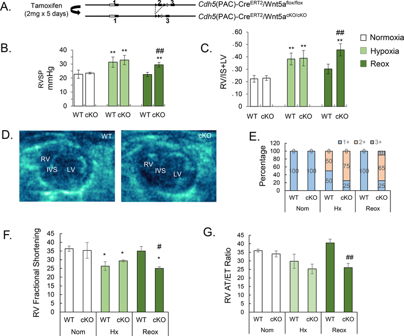
Isolation of pericytes and PMVECs from healthy donor and PAH lungs was carried out with 3G5 or CD31 antibody-conjugated magnetic beads. Wnt expression profile of PMVECs was documented via quantitative polymerase chain reaction with a Wnt primer library. Exosome purification from PMVEC media was carried out with the ExoTIC device. Hemodynamic profile, right ventricular function, and pulmonary vascular morphometry were obtained in a conditional endothelium-specific Wnt5a knockout ( Wnt5a) mouse model under normoxia, chronic hypoxia, and hypoxia recovery.
Quantification of Wnt ligand expression in healthy PMVECs cocultured with pericytes demonstrated a 35-fold increase in Wnt5a, a known Wnt/planar cell polarity ligand. This Wnt5a spike was not seen in PAH PMVECs, which correlated with an inability to recruit pericytes in Matrigel coculture assays. Exosomes purified from media demonstrated an increase in Wnt5a content when healthy PMVECs were cocultured with pericytes, a finding that was not observed in exosomes of PAH PMVECs. Furthermore, the addition of either recombinant Wnt5a or purified healthy PMVEC exosomes increased pericyte recruitment to PAH PMVECs in coculture studies. Although no differences were noted in normoxia and chronic hypoxia, Wnt5a mice demonstrated persistent pulmonary hypertension and right ventricular failure 4 weeks after recovery from chronic hypoxia, which correlated with significant reduction, muscularization, and decreased pericyte coverage of microvessels.
We identify Wnt5a as a key mediator for the establishment of pulmonary endothelium-pericyte interactions, and its loss could contribute to PAH by reducing the viability of newly formed vessels. We speculate that therapies that mimic or restore Wnt5a production could help prevent loss of small vessels in PAH.
肺动脉高压(PAH)是一种危及生命的肺循环疾病,与微血管的丧失和再生障碍有关。肺微血管周细胞覆盖减少是 PAH 的病理特征之一,部分原因是周细胞无法对肺微血管内皮细胞(PMVEC)发出的信号做出反应。我们已经表明,Wnt/平面细胞极性通路的激活是周细胞募集所必需的,但 PMVEC 产生和释放特定的 Wnt 配体是否负责周细胞中的 Wnt/平面细胞极性激活尚不清楚。
使用 3G5 或 CD31 抗体偶联磁珠从健康供体和 PAH 肺中分离周细胞和 PMVEC。通过定量聚合酶链反应和 Wnt 引物文库记录 PMVEC 的 Wnt 表达谱。使用 ExoTIC 设备从 PMVEC 培养基中纯化外泌体。在正常氧、慢性缺氧和缺氧恢复条件下,在条件性内皮细胞特异性 Wnt5a 敲除(Wnt5a)小鼠模型中获得血流动力学特征、右心室功能和肺血管形态计量学。
在与周细胞共培养的健康 PMVEC 中定量检测 Wnt 配体表达显示 Wnt5a 增加了 35 倍,Wnt5a 是一种已知的 Wnt/平面细胞极性配体。这种 Wnt5a 峰在 PAH PMVEC 中未见,这与在 Matrigel 共培养测定中无法募集周细胞有关。当健康 PMVEC 与周细胞共培养时,从培养基中纯化的外泌体显示 Wnt5a 含量增加,但在 PAH PMVEC 的外泌体中没有观察到这种情况。此外,在共培养研究中,添加重组 Wnt5a 或纯化的健康 PMVEC 外泌体均可增加 PAH PMVEC 中周细胞的募集。虽然在正常氧和慢性缺氧条件下没有差异,但 Wnt5a 小鼠在慢性缺氧恢复后 4 周仍表现出持续性肺动脉高压和右心衰竭,这与微血管的显著减少、肌化和周细胞覆盖减少有关。
我们将 Wnt5a 确定为建立肺内皮细胞-周细胞相互作用的关键介质,其缺失可能通过降低新形成血管的活力而导致 PAH。我们推测,模拟或恢复 Wnt5a 产生的治疗方法可能有助于防止 PAH 中小血管的丧失。