Katari Yeshaswini, Srinivasan Rangalakshmi, Arvind Priyadarshini, Hiremathada Sahajananda
Department of Anesthesiology and Critical Care, Rajarajeswari Medical College and Hospital, Bangalore, Karnataka, India.
Indian J Crit Care Med. 2018 Nov;22(11):781-788. doi: 10.4103/ijccm.IJCCM_394_18.
Muscular atrophy is the universal feature in patients with ICUAW. Muscles of the lower limb are more prone to early atrophy. Measurement of fat thickness is used to assess malnutrition. This study was designed to evaluate if, subcutaneous fat also reduces along with muscle thickness and if it can be reliably used as an indicator of nutritional assessment in critically ill patients using point of care ultrasound.
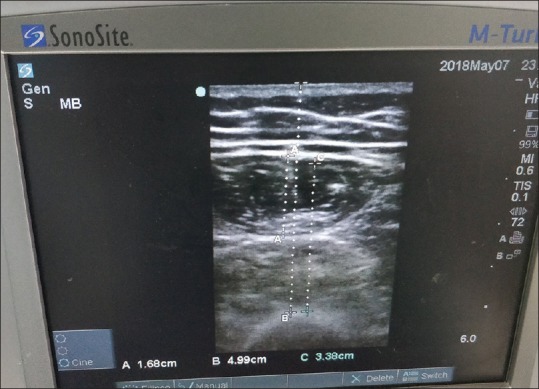
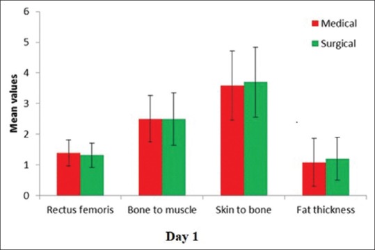
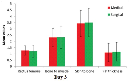
An observational clinical study of 100 patients admitted to multidisciplinary intensive care units (ICUs). Total anterior thigh thickness, thickness of the rectus femoris muscle, fat thickness, and the combined thickness of vastus intermedius and rectus femoris were taken on day 1, 3, and 7 of ICU admission.
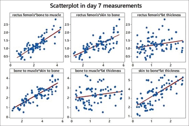
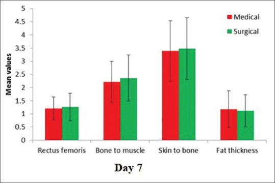
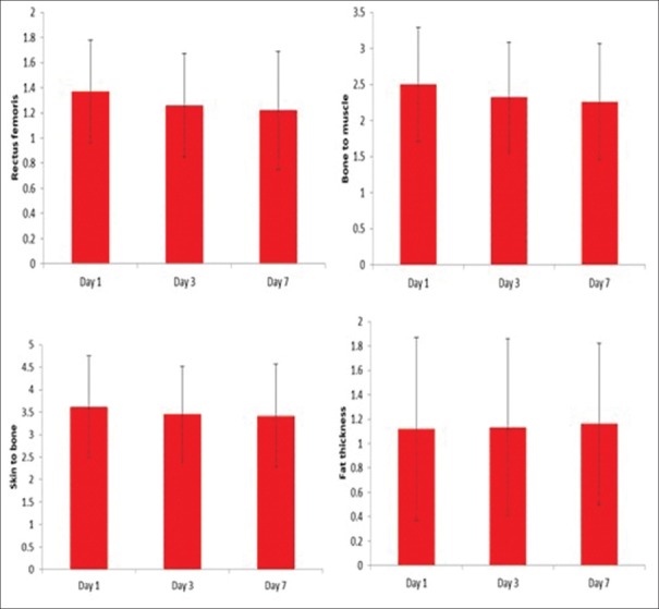
There was progressive loss of muscle mass from day 1 to day 7. Muscle loss was not only limited to rectus femoris, but vastus intermedius also showed a significant decrease as indicated by the bone to muscle measurement. Skin to bone measurement which includes both muscle and fat compartment showed a decline.
There is potential utility of ultrasound for early detection and probable corrective measures to prevent ICUAW. The rectus femoris thickness, skin to bone, and bone to muscle thickness show statistically significant difference on day 3, day 7 compared to day 1. Fat layer did not show statistically significant decrease.
肌肉萎缩是重症监护病房获得性肌无力(ICUAW)患者的普遍特征。下肢肌肉更容易早期萎缩。测量脂肪厚度用于评估营养不良状况。本研究旨在评估皮下脂肪是否也会随着肌肉厚度的减少而减少,以及在危重症患者中,使用床旁超声它是否能可靠地用作营养评估指标。
对100例入住多学科重症监护病房(ICU)的患者进行一项观察性临床研究。在入住ICU的第1天、第3天和第7天测量大腿前侧总厚度、股直肌厚度、脂肪厚度以及股中间肌和股直肌的联合厚度。
从第1天到第7天肌肉量逐渐减少。肌肉减少不仅限于股直肌,股中间肌也显示出显著减少,这由骨与肌肉的测量结果表明。包括肌肉和脂肪层的皮肤与骨的测量结果显示下降。
超声对于早期检测ICUAW以及采取可能的纠正措施具有潜在作用。股直肌厚度、皮肤与骨厚度以及骨与肌肉厚度在第3天、第7天与第1天相比有统计学显著差异。脂肪层未显示出统计学显著减少。