National Health Commission Key Laboratory of Hearing Medicine (Fudan University), Department of Otology and Skull Base Surgery, Shanghai, China; Hospital of Xuzhou Medical University, Department of Otolaryngology, Xuzhou, China.
National Health Commission Key Laboratory of Hearing Medicine (Fudan University), Department of Otology and Skull Base Surgery, Shanghai, China.
Braz J Otorhinolaryngol. 2020 Mar-Apr;86(2):165-173. doi: 10.1016/j.bjorl.2018.10.014. Epub 2018 Dec 20.
Meniere's disease is associated with impaired hearing, tinnitus, vertigo, and aural fullness. Many anatomical studies have suggested idiopathic endolymphatic hydrops as the pathological basis of Meniere's disease, which now can be visualized by using gadolinium -enhanced magnetic resonance imaging of the inner ear.
To investigate the development of endolymphatic hydrops in Meniere's disease by monitoring the vestibules and cochleae of affected patients.
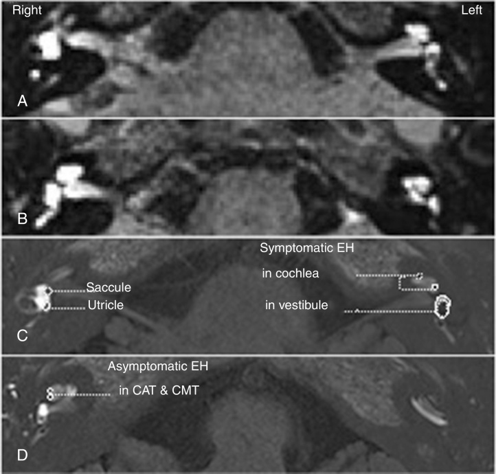
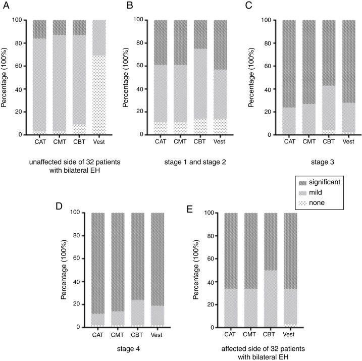
Inner ears of 178 patients with definite unilateral Meniere's disease diagnosis were visualized by 3-dimensional fluid-attenuated inversion recovery and three-dimensional real inversion recovery magnetic resonance imaging following bilateral gadolinium intratympanic injection. The scans were used to evaluate the presence and degree of endolymphatic hydrops in the vestibules and cochlear structures, including the cochlear apical turn, the cochlear middle turn, and the cochlear basal turn. The correlation of endolymphatic hydrops occurrence between the various parts of the inner ear was determined.
Symptomatic endolymphatic hydrops was detected on the affected side in all patients, whereas asymptomatic endolymphatic hydrops was detected on the unaffected contra-lateral side in 32 patients (18.0%). On the affected side, the cochlear apical turn and the cochlear middle turn demonstrated significantly higher rates of endolymphatic hydrops than the cochlear basal turn and the vestibule. The severity of endolymphatic hydrops gradually decreased from the cochlear apical turn to the cochlear basal turn. On the contra lateral side, the incidence and degree of the detected asymptomatic endolymphatic hydrops were significantly greater in the cochleae than in the vestibules (p<0.05), with no significant difference detected between the cochlear turns.
Progression of endolymphatic hydrops appears to be directional, initiated in the cochlea. The order of endolymphatic hydrops severity gradually decreases from the cochlear apical turn to the cochlear basal turn and then to the vestibule. Endolymphatic hydrops in the vestibule is associated with symptomatic Meniere's disease.
梅尼埃病与听力下降、耳鸣、眩晕和耳部饱满感有关。许多解剖学研究表明特发性内淋巴积水是梅尼埃病的病理基础,现在可以通过使用钆增强内耳磁共振成像来观察到。
通过监测受影响患者的前庭和耳蜗,研究梅尼埃病内淋巴积水的发展。
对 178 例单侧梅尼埃病诊断明确的患者进行双侧鼓室内钆注射后,采用三维液体衰减反转恢复和三维真实反转恢复磁共振成像对内耳进行可视化。扫描用于评估前庭和耳蜗结构(包括耳蜗顶回、耳蜗中回和耳蜗底回)内内淋巴积水的存在和程度。确定内耳各部位内淋巴积水发生的相关性。
所有患者均在受影响侧检测到症状性内淋巴积水,而在 32 例(18.0%)非受影响对侧检测到无症状性内淋巴积水。在受影响侧,耳蜗顶回和耳蜗中回的内淋巴积水发生率明显高于耳蜗底回和前庭。内淋巴积水的严重程度从耳蜗顶回逐渐下降到耳蜗底回。在对侧,检测到的无症状性内淋巴积水的发生率和程度在耳蜗中明显大于前庭(p<0.05),但耳蜗各回之间无显著差异。
内淋巴积水的进展似乎是定向的,始于耳蜗。内淋巴积水严重程度的顺序从耳蜗顶回逐渐下降到耳蜗底回,然后到前庭。前庭内淋巴积水与有症状的梅尼埃病有关。