• 文献检索
  • 文档翻译
  • 深度研究
  • 学术资讯
  • Suppr Zotero 插件Zotero 插件
  • 邀请有礼
  • 套餐&价格
  • 历史记录
应用&插件
Suppr Zotero 插件Zotero 插件浏览器插件Mac 客户端Windows 客户端微信小程序
定价
高级版会员购买积分包购买API积分包
服务
文献检索文档翻译深度研究API 文档MCP 服务
关于我们
关于 Suppr公司介绍联系我们用户协议隐私条款
关注我们

Suppr 超能文献

核心技术专利:CN118964589B侵权必究
粤ICP备2023148730 号-1Suppr @ 2026

文献检索

告别复杂PubMed语法,用中文像聊天一样搜索,搜遍4000万医学文献。AI智能推荐,让科研检索更轻松。

立即免费搜索

文件翻译

保留排版,准确专业,支持PDF/Word/PPT等文件格式,支持 12+语言互译。

免费翻译文档

深度研究

AI帮你快速写综述,25分钟生成高质量综述,智能提取关键信息,辅助科研写作。

立即免费体验

用于下颌第三磨牙深度阻生拔除术的计算机辅助设计/计算机辅助制造切割导板

Computer-Aided Design/Computer-Aided Manufacturing Cutting Guides for Odontectomy of Deeply Impacted Mandibular Third Molars.

作者信息

Ahmed Mamdouhh, Salah Mariam Kamel, Khairy Nesrine

机构信息

Faculty of Dentistry, Cairo University, Oral and Maxillofacial Department 11 Al Saraya, Al Manial, Giza Governorate 11553, Egypt.

出版信息

Open Access Maced J Med Sci. 2018 Dec 19;6(12):2395-2401. doi: 10.3889/oamjms.2018.371. eCollection 2018 Dec 20.

DOI:10.3889/oamjms.2018.371
PMID:30607200
原文链接:https://pmc.ncbi.nlm.nih.gov/articles/PMC6311491/
Abstract

AIM

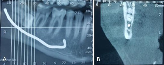
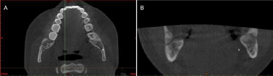
To evaluate a new technique for surgical removal of deeply impacted mandibular third molars (DIMTM), using computer-guided cutting guide to maintain inferior alveolar nerve (IAN) integrity and the covering buccal bone.

PATIENTS AND METHODS

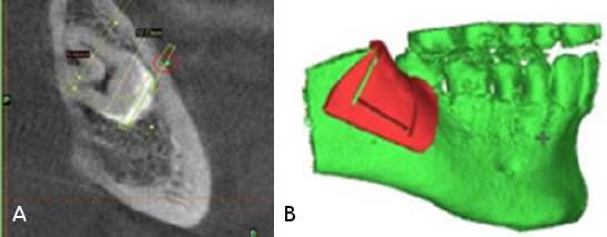
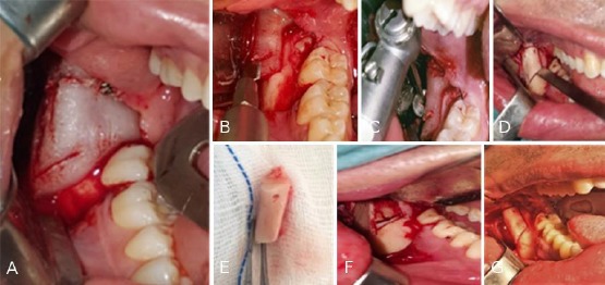
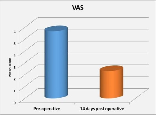
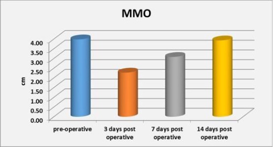
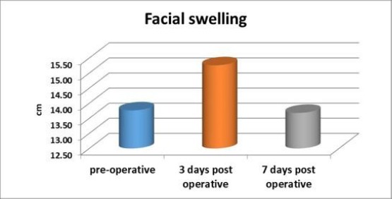
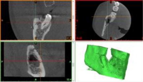
Eighteen cases indicated for removal of DIMTM. Cone-beam Computed Tomography (CBCTs) used to determine the tooth's relation to the IAN. Computer-guided software used for fabrication of surgical cutting guide stent to expose the impacted tooth and repositioning of bone after odontectomy without fixation. Clinical assessment included a neurosensory deficit of IAN, pain using a visual analogue scale (VAS), facial swelling, and maximal mouth opening (MMO). CBCTs were taken immediately and six months postoperatively to evaluate position and healing of bone.

RESULTS

None of the patients showed a permanent neurological deficit of IAN while all patients showed normal parameters of pain, facial swelling and MMO.

CONCLUSION

this technique has shown the accurate determination of the bony window cuts with subsequent preservation of IAN and external oblique ridge.

摘要

目的

评估一种使用计算机引导切割导板来维持下牙槽神经(IAN)完整性和颊侧覆盖骨的手术切除深部下颌阻生第三磨牙(DIMTM)的新技术。

患者与方法

18例需切除DIMTM的病例。使用锥形束计算机断层扫描(CBCT)确定牙齿与IAN的关系。使用计算机引导软件制作手术切割导板支架,以暴露阻生牙并在拔牙后重新定位骨组织,无需固定。临床评估包括IAN的神经感觉功能缺损、使用视觉模拟量表(VAS)评估的疼痛、面部肿胀和最大开口度(MMO)。在术后即刻和术后6个月拍摄CBCT,以评估骨组织的位置和愈合情况。

结果

所有患者均未出现IAN永久性神经功能缺损,且所有患者的疼痛、面部肿胀和MMO参数均正常。

结论

该技术已显示出能准确确定骨窗切口,随后可保留IAN和外斜线。

https://cdn.ncbi.nlm.nih.gov/pmc/blobs/a780/6311491/3cf46f79d261/OAMJMS-6-2395-g008.jpg
https://cdn.ncbi.nlm.nih.gov/pmc/blobs/a780/6311491/b054ad73fca9/OAMJMS-6-2395-g001.jpg
https://cdn.ncbi.nlm.nih.gov/pmc/blobs/a780/6311491/6a427d53e59a/OAMJMS-6-2395-g002.jpg
https://cdn.ncbi.nlm.nih.gov/pmc/blobs/a780/6311491/a56f839416cd/OAMJMS-6-2395-g003.jpg
https://cdn.ncbi.nlm.nih.gov/pmc/blobs/a780/6311491/b4d6793ff4b5/OAMJMS-6-2395-g004.jpg
https://cdn.ncbi.nlm.nih.gov/pmc/blobs/a780/6311491/562f0ea9db29/OAMJMS-6-2395-g005.jpg
https://cdn.ncbi.nlm.nih.gov/pmc/blobs/a780/6311491/3376b70ca4d1/OAMJMS-6-2395-g006.jpg
https://cdn.ncbi.nlm.nih.gov/pmc/blobs/a780/6311491/8e9a571c5b62/OAMJMS-6-2395-g007.jpg
https://cdn.ncbi.nlm.nih.gov/pmc/blobs/a780/6311491/3cf46f79d261/OAMJMS-6-2395-g008.jpg

相似文献

1
Computer-Aided Design/Computer-Aided Manufacturing Cutting Guides for Odontectomy of Deeply Impacted Mandibular Third Molars.用于下颌第三磨牙深度阻生拔除术的计算机辅助设计/计算机辅助制造切割导板
Open Access Maced J Med Sci. 2018 Dec 19;6(12):2395-2401. doi: 10.3889/oamjms.2018.371. eCollection 2018 Dec 20.
2
Protocol for Diagnostic Test Accuracy Study: Evaluation of Cone Beam Computed Tomography (CBCT) in Prediction of Inferior Alveolar Nerve Injury as Compared to Orthopantomography (OPG) Secondary to Surgical Removal of Impacted Mandibular Third Molars.诊断试验准确性研究方案:与曲面体层摄影术(OPG)相比,锥形束计算机断层扫描(CBCT)在预测因拔除下颌阻生第三磨牙继发下牙槽神经损伤中的评估
Cureus. 2024 Aug 14;16(8):e66864. doi: 10.7759/cureus.66864. eCollection 2024 Aug.
3
Removal of impacted mandibular third molars using an inward fragmentation technique (IFT) - Method and first results.使用向内分割技术(IFT)拔除下颌阻生第三磨牙 - 方法及初步结果。
J Craniomaxillofac Surg. 2014 Apr;42(3):213-9. doi: 10.1016/j.jcms.2013.05.003. Epub 2013 Jul 11.
4
Assessment of the relationship between impacted mandibular third molars and inferior alveolar nerve with dental 3-dimensional computed tomography.利用牙科三维计算机断层扫描评估下颌阻生第三磨牙与下牙槽神经之间的关系。
J Oral Maxillofac Surg. 2009 Dec;67(12):2587-91. doi: 10.1016/j.joms.2009.07.017.
5
Evaluation of Outcome Following Coronectomy for the Management of Mandibular Third Molars in Close Proximity to Inferior Alveolar Nerve.下颌第三磨牙紧邻下牙槽神经时采用截冠术治疗后的疗效评估
J Clin Diagn Res. 2016 Aug;10(8):ZC57-62. doi: 10.7860/JCDR/2016/20991.8273. Epub 2016 Aug 1.
6
New quantitative classification of the anatomical relationship between impacted third molars and the inferior alveolar nerve.阻生第三磨牙与下牙槽神经解剖关系的新定量分类
BMC Med Imaging. 2015 Dec 7;15:59. doi: 10.1186/s12880-015-0101-0.
7
Position of the impacted third molar in relation to the mandibular canal. Diagnostic accuracy of cone beam computed tomography compared with panoramic radiography.阻生第三磨牙相对于下颌管的位置。锥束计算机断层扫描与全景X线摄影的诊断准确性比较。
Int J Oral Maxillofac Surg. 2009 Sep;38(9):964-71. doi: 10.1016/j.ijom.2009.06.007. Epub 2009 Jul 28.
8
Factors that are associated with injury to the inferior alveolar nerve in high-risk patients after removal of third molars.拔除第三磨牙后高危患者下牙槽神经损伤的相关因素。
Br J Oral Maxillofac Surg. 2013 Dec;51(8):868-73. doi: 10.1016/j.bjoms.2013.08.007. Epub 2013 Sep 3.
9
Clinical relevance of cone beam computed tomography in mandibular third molar removal: A multicentre, randomised, controlled trial.锥形束计算机断层扫描在下颌第三磨牙拔除术中的临床相关性:一项多中心、随机、对照试验。
J Craniomaxillofac Surg. 2015 Dec;43(10):2158-67. doi: 10.1016/j.jcms.2015.10.009. Epub 2015 Oct 23.
10
Inferior alveolar nerve sensory disturbance after impacted mandibular third molar evaluation using cone beam computed tomography and panoramic radiography: a pilot study.使用锥形束计算机断层扫描和全景X线摄影评估下颌阻生第三磨牙后下牙槽神经感觉障碍:一项初步研究。
J Oral Maxillofac Surg. 2012 Oct;70(10):2264-70. doi: 10.1016/j.joms.2012.04.015. Epub 2012 Jun 16.

引用本文的文献

1
Three-dimensional reconstruction of cone beam CT scan in the planning of the surgery of mandibular impacted third molars.锥形束CT扫描三维重建在下颌阻生第三磨牙手术规划中的应用
Natl J Maxillofac Surg. 2025 Jan-Apr;16(1):33-38. doi: 10.4103/njms.njms_135_22. Epub 2025 Apr 28.
2
Association of the Inferior Alveolar Nerve Position and Nerve Injury: A Systematic Review and Meta-Analysis.下牙槽神经位置与神经损伤的关联:一项系统评价与Meta分析
Healthcare (Basel). 2022 Sep 16;10(9):1782. doi: 10.3390/healthcare10091782.
3
The bone lid technique in lateral sinus lift: a systematic review and meta-analysis.

本文引用的文献

1
Radiographic features of anatomic relationship between impacted third molar and inferior alveolar canal on coronal CBCT images: risk factors for nerve injury after tooth extraction.锥形束计算机断层扫描(CBCT)冠状位图像上阻生第三磨牙与下牙槽神经管解剖关系的影像学特征:拔牙后神经损伤的危险因素
Arch Med Sci. 2018 Apr;14(3):532-540. doi: 10.5114/aoms.2016.58842. Epub 2016 Mar 23.
2
Removal of Deeply Impacted Mandibular Molars by Sagittal Split Osteotomy.经矢状劈开截骨术拔除深度阻生下颌磨牙
Case Rep Dent. 2016;2016:1902089. doi: 10.1155/2016/1902089. Epub 2016 Jun 27.
3
New quantitative classification of the anatomical relationship between impacted third molars and the inferior alveolar nerve.
骨瓣技术在侧窦提升术中的应用:系统评价和荟萃分析。
Int J Implant Dent. 2022 Aug 29;8(1):33. doi: 10.1186/s40729-022-00433-3.
4
The Bone Lid Technique in Oral and Maxillofacial Surgery: A Scoping Review.口腔颌面外科中的骨盖技术:一项范围综述
J Clin Med. 2022 Jun 24;11(13):3667. doi: 10.3390/jcm11133667.
阻生第三磨牙与下牙槽神经解剖关系的新定量分类
BMC Med Imaging. 2015 Dec 7;15:59. doi: 10.1186/s12880-015-0101-0.
4
A multivariate statistical analysis on variables affecting inferior alveolar nerve damage during third molar surgery.对影响第三磨牙手术中下牙槽神经损伤的变量进行的多变量统计分析。
Br Dent J. 2015 Aug 28;219(4):E3. doi: 10.1038/sj.bdj.2015.661.
5
Impacted Mandibular Third Molars: Review of Literature and a Proposal of a Combined Clinical and Radiological Classification.下颌阻生第三磨牙:文献综述及临床与影像学联合分类建议
Ann Med Health Sci Res. 2015 Jul-Aug;5(4):229-34. doi: 10.4103/2141-9248.160177.
6
Coronectomy as a surgical approach to impacted mandibular third molars: a systematic review.冠切除术作为下颌阻生第三磨牙的一种手术方法:一项系统评价
Head Face Med. 2015 Apr 10;11:9. doi: 10.1186/s13005-015-0068-7.
7
Inferior alveolar nerve injury after mandibular third molar extraction: a literature review.下颌第三磨牙拔除术后下牙槽神经损伤:文献综述
J Oral Maxillofac Res. 2014 Dec 29;5(4):e1. doi: 10.5037/jomr.2014.5401. eCollection 2014 Oct-Dec.
8
Virtual surgical planning in craniofacial surgery.颅颌面外科中的虚拟手术规划。
Semin Plast Surg. 2014 Aug;28(3):150-8. doi: 10.1055/s-0034-1384811.
9
Mandibular third molar impaction: review of literature and a proposal of a classification.下颌第三磨牙阻生:文献综述及分类建议
J Oral Maxillofac Res. 2013 Jul 1;4(2):e1. doi: 10.5037/jomr.2013.4201.
10
The relative risk of neurosensory deficit following removal of mandibular third molar teeth: the influence of radiography and surgical technique.下颌第三磨牙拔除后发生神经感觉缺陷的相对风险:影像学检查和手术技术的影响。
Oral Surg Oral Med Oral Pathol Oral Radiol. 2013 Jan;115(1):18-24. doi: 10.1016/j.oooo.2012.03.017. Epub 2012 Aug 22.