Ancora Daniele, Qiu Lina, Zacharakis Giannis, Spinelli Lorenzo, Torricelli Alessandro, Pifferi Antonio
Institute of Electronic Structure and Laser, Foundation for Research and Technology - Hellas, Heraklion, Greece.
Department of Materials Science and Technology, University of Crete, Heraklion, Greece.
Biomed Opt Express. 2018 Aug 6;9(9):4094-4112. doi: 10.1364/BOE.9.004094. eCollection 2018 Sep 1.
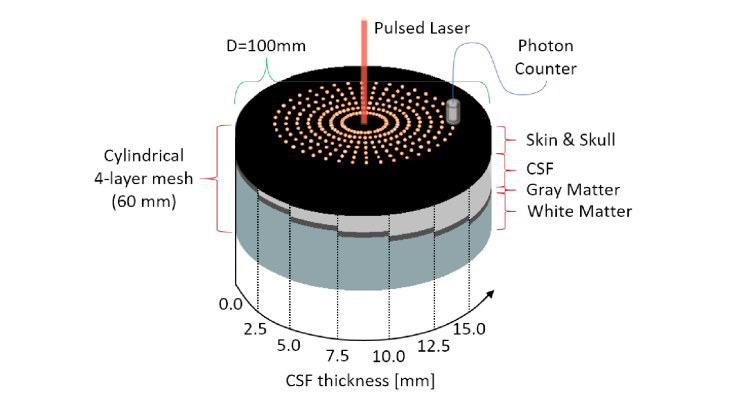
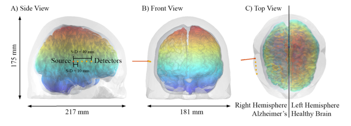
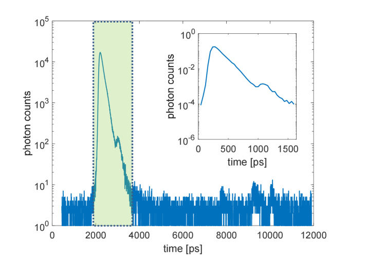
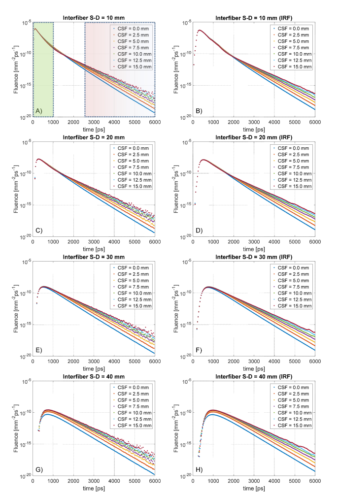
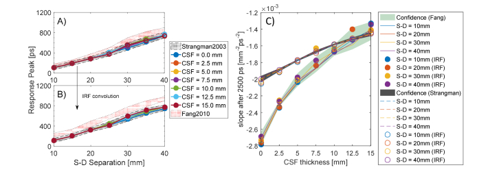
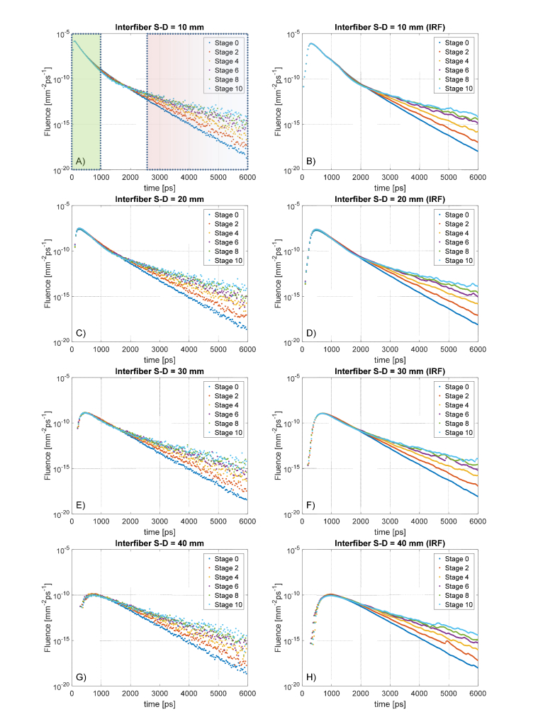
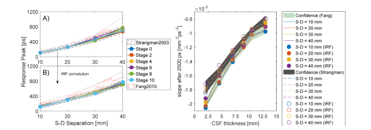
Dementia disorders are increasingly becoming sources of a broad range of problems, strongly interfering with the normal daily tasks of a growing number of individuals. Such neurodegenerative diseases are often accompanied with progressive brain atrophy that, at late stages, leads to drastically reduced brain dimensions. Currently, this structural change could be followed with X-ray computed tomography (XCT) or magnetic resonance imaging (MRI), but they share numerous disadvantages in terms of usability, invasiveness and costs. In this work, we aim to retrieve information concerning the brain-atrophy stage and its evolution, proposing a novel approach based on non-invasive time-resolved near infra-red (tr-NIR) measurements. For this purpose, we created a set of virtual human-head atlases in which we eroded the brain as it would happen in a clinical brain-atrophy progression. These realistic meshes were used to simulate a longitudinal tr-NIR study, investigating the effects of an increased amount of cerebral spinal fluid (CSF) in the photon diffusion. The analysis of late photons in the time-resolved reflectance curve-obtained via accurate Monte Carlo simulations-exhibited peculiar slope-changes upon CSF layer increase. The visibility of the effect under several measurement conditions suggested good sensitivity to CSF variation, even in the case of real measurement and under different geometrical models. The robustness of the results might promote the technique as a potential indicator of the dementia progression, relying only on fast and non-invasive optical observations.
痴呆症正日益成为一系列广泛问题的根源,严重干扰着越来越多个人的正常日常活动。此类神经退行性疾病通常伴有进行性脑萎缩,在疾病后期会导致脑尺寸大幅减小。目前,可以通过X射线计算机断层扫描(XCT)或磁共振成像(MRI)来跟踪这种结构变化,但它们在可用性、侵入性和成本方面存在诸多缺点。在这项工作中,我们旨在获取有关脑萎缩阶段及其演变的信息,提出一种基于非侵入性时间分辨近红外(tr-NIR)测量的新方法。为此,我们创建了一组虚拟人头图谱,在其中按照临床脑萎缩进展的情况对大脑进行侵蚀。这些逼真的网格模型用于模拟一项纵向tr-NIR研究,探究脑脊液(CSF)量增加对光子扩散的影响。通过精确的蒙特卡罗模拟获得的时间分辨反射率曲线中晚期光子的分析表明,随着CSF层增加,曲线呈现出特殊的斜率变化。在几种测量条件下该效应的可见性表明,即使在实际测量以及不同几何模型的情况下,该方法对CSF变化也具有良好的敏感性。结果的稳健性可能会促使该技术成为痴呆症进展的潜在指标,仅依靠快速且非侵入性的光学观测即可。