Li Ying, Struebing Felix L, Wang Jiaxing, King Rebecca, Geisert Eldon E
Department of Ophthalmology, Emory University, Atlanta, GA, United States.
Center for Neuropathology and Prion Research, Ludwig Maximilian University of Munich, Munich, Germany.
Front Genet. 2018 Dec 18;9:633. doi: 10.3389/fgene.2018.00633. eCollection 2018.
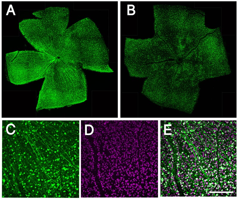
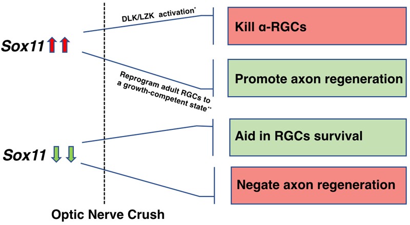
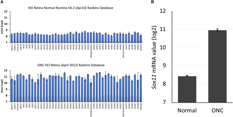
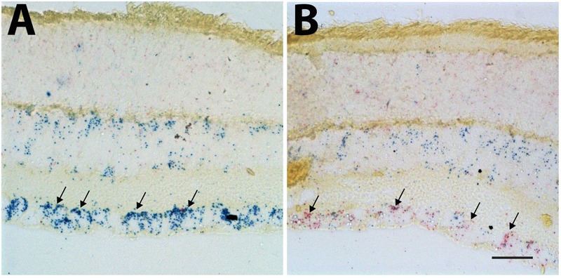
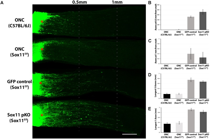
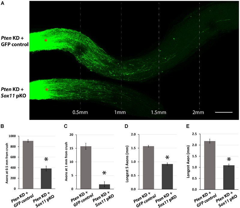
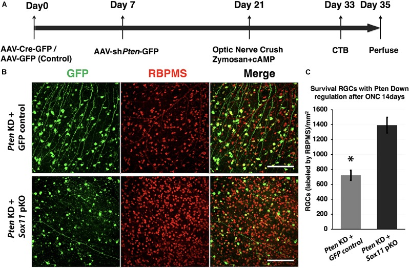
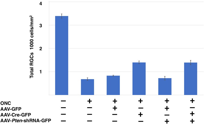
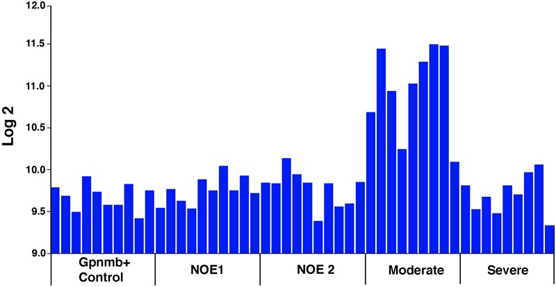
The present study examines the role of in the initial response of retinal ganglion cells (RGCs) to axon damage and in optic nerve regeneration in mouse. Markers of retinal injury were identified using the normal retina database and optic nerve crush (ONC) database on GeneNetwork (www.genenetwork.org). One gene, , was highly upregulated following ONC. We examined the role of this transcription factor, , following ONC and optic nerve regeneration in mice. hybridization was performed using the Affymetrix 2-plex Quantigene View RNA Hybridization Tissue Assay System. was partially knocked out by intravitreal injection of AAV2-CMV-Cre-GFP in mice. Optic nerve regeneration model used knockdown. Mice were perfused and the retinas and optic nerves were dissected and examined for RGC survival and axon growth. was dramatically upregulated in the retina following ONC injury. The level of message increased by approximately eightfold 2 days after ONC. hybridization demonstrated low-level message in RGCs and cells in the inner nuclear layer in the normal retina as well as a profound increase in message within the ganglion cells following ONC. In retinas, partially knocking out significantly increased RGC survival after ONC as compared to the AAV2-CMV-GFP control group; however, it had little effect on the ability of axon regeneration. Combinatorial downregulation of both and resulted in a significant increase in RGC survival as compared to knockdown only. When was knocked down there was a remarkable increase in the number and the length of regenerating axons. Partially knocking out in combination with deletion resulted in a fewer regenerating axons. Taken together, these data demonstrate that is involved in the initial response of the retina to injury, playing a role in the early attempts of axon regeneration and neuronal survival. Downregulation of aids in RGC survival following injury of optic nerve axons, while a partial knockout of negates the axon regeneration stimulated by knockdown.
本研究探讨了[具体基因名称]在小鼠视网膜神经节细胞(RGCs)对轴突损伤的初始反应以及视神经再生中的作用。利用GeneNetwork(www.genenetwork.org)上的正常视网膜数据库和视神经挤压(ONC)数据库确定了视网膜损伤的标志物。一个基因[具体基因名称]在ONC后高度上调。我们研究了这种转录因子[具体基因名称]在小鼠ONC和视神经再生后的作用。使用Affymetrix 2重plex Quantigene View RNA杂交组织检测系统进行杂交。通过向[具体基因名称]小鼠玻璃体内注射AAV2-CMV-Cre-GFP部分敲除[具体基因名称]。视神经再生模型采用[具体基因名称]敲低。对小鼠进行灌注,解剖视网膜和视神经,检查RGC存活和轴突生长情况。ONC损伤后,[具体基因名称]在视网膜中显著上调。ONC后2天,[具体基因名称]的信使水平增加了约8倍。杂交显示正常视网膜中RGCs和内核层细胞中的[具体基因名称]信使水平较低,而ONC后神经节细胞内的[具体基因名称]信使水平显著增加。在[具体基因名称]视网膜中,与AAV2-CMV-GFP对照组相比,部分敲除[具体基因名称]显著增加了ONC后RGC的存活;然而,它对轴突再生能力影响不大。与仅敲低[具体基因名称]相比,同时下调[具体基因名称]和[另一具体基因名称]导致RGC存活显著增加。当敲低[具体基因名称]时,再生轴突的数量和长度显著增加。部分敲除[具体基因名称]与缺失[另一具体基因名称]相结合导致再生轴突减少。综上所述,这些数据表明[具体基因名称]参与视网膜对损伤的初始反应,在轴突再生和神经元存活的早期尝试中发挥作用。下调[具体基因名称]有助于视神经轴突损伤后RGC的存活,而部分敲除[具体基因名称]则消除了由[具体基因名称]敲低刺激的轴突再生。
需注意,原文中部分具体基因名称未给出具体内容,所以译文里用[具体基因名称]等进行了标注。