Al-Sharydah Abdulaziz Mohammad, Al-Arfaj Hussain Khalid, Saleh Al-Muhaish Husam, Al-Suhaibani Sari Saleh, Al-Aftan Mohammad Saad, Almedallah Dana Khaled, Al-Abdulwahhab Abdulrhman Hamad, Al-Hedaithy Abdullah Abdulaziz, Al-Jubran Saeed Ahmad
Radiology Department, Imam Abdulrahman Bin Faisal University, King Fahd Hospital of the University, Al-Khobar City, Eastern Province, Saudi Arabia.
Medical Imaging Department, King Fahad Specialist Hospital, Dammam City, Eastern Province, Saudi Arabia.
Eur J Radiol Open. 2019 Jan 4;6:49-55. doi: 10.1016/j.ejro.2018.12.004. eCollection 2019.
Classifying brain tumors is challenging, but recently developed imaging techniques offer the opportunity for neuroradiologists and neurosurgeons to diagnose, differentiate, and manage different types of brain tumors. Such advances will be reflected in improvements in patients' life expectancy and quality of life. Among the newest techniques, the apparent diffusion coefficient (ADC), which tracks the rate of microscopic water diffusion within tissues, has become a focus of investigation. Recently, ADC has been used as a preoperative diffusion-weighted magnetic resonance imaging (MRI) parameter that facilitates tumor diagnosis and grading. Here, we aimed to determine the ADC cutoff values for pediatric brain tumors (PBTs) categorized according to the World Health Organization (WHO) classification of brain tumors.
We retrospectively reviewed 80 cases, and assessed them based on their MRI-derived ADC. These results were compared with those of WHO classification-based histopathology.
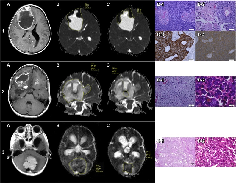
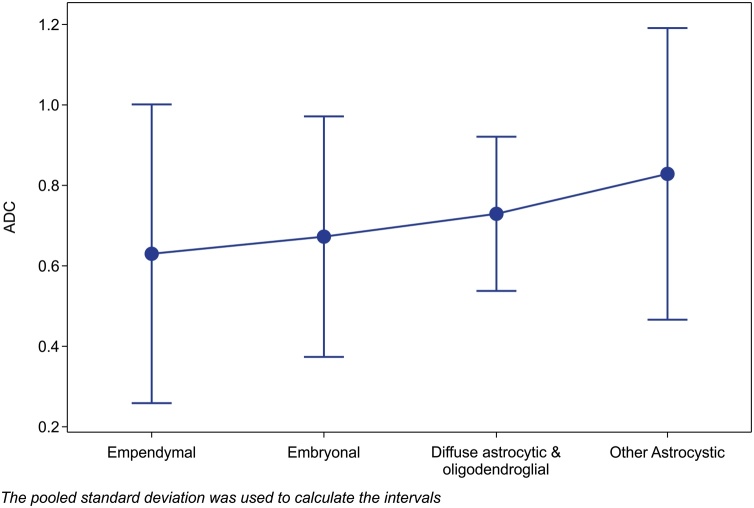
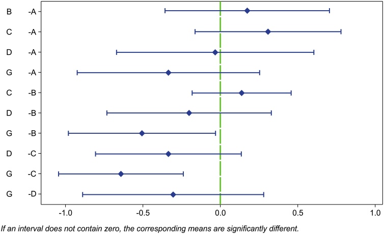
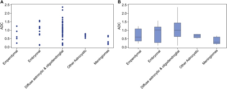
Whole-lesion ADC values ranged 0.225-1.240 × 10 mm/s for ependymal tumors, 0.107-1.571 × 10 mm/s for embryonal tumors, 0.1065-2.37801 × 10 mm/s for diffuse astrocytic and oligodendroglial tumors, 0.5220-0.7840 × 10 mm/s for other astrocytic tumors, and 0.1530-0.8160 × 10 mm/s for meningiomas. These findings revealed the usefulness of ADC in the differential diagnosis of PBT, as it was able to discriminate between five types of PBTs.
The application of an ADC diagnostic criterion would reduce the need for spectroscopic analysis. However, further research is needed to implement ADC in the differential diagnosis of PBT.
脑肿瘤的分类具有挑战性,但最近开发的成像技术为神经放射科医生和神经外科医生诊断、区分和管理不同类型的脑肿瘤提供了机会。这些进展将反映在患者预期寿命和生活质量的提高上。在最新技术中,表观扩散系数(ADC)可追踪组织内微观水扩散速率,已成为研究焦点。最近,ADC已被用作术前扩散加权磁共振成像(MRI)参数,有助于肿瘤诊断和分级。在此,我们旨在确定根据世界卫生组织(WHO)脑肿瘤分类法分类的小儿脑肿瘤(PBT)的ADC临界值。
我们回顾性分析了80例病例,并根据MRI得出的ADC对其进行评估。将这些结果与基于WHO分类的组织病理学结果进行比较。
室管膜瘤全瘤ADC值范围为0.225 - 1.240×10⁻³mm²/s,胚胎性肿瘤为0.107 - 1.571×10⁻³mm²/s,弥漫性星形细胞和少突胶质细胞瘤为0.1065 - 2.37801×10⁻³mm²/s,其他星形细胞瘤为0.5220 - 0.7840×10⁻³mm²/s,脑膜瘤为0.1530 - 0.8160×10⁻³mm²/s。这些发现揭示了ADC在PBT鉴别诊断中的有用性,因为它能够区分五种类型的PBT。
应用ADC诊断标准将减少对光谱分析的需求。然而,需要进一步研究以在PBT的鉴别诊断中应用ADC。