• 文献检索
  • 文档翻译
  • 深度研究
  • 学术资讯
  • Suppr Zotero 插件Zotero 插件
  • 邀请有礼
  • 套餐&价格
  • 历史记录
应用&插件
Suppr Zotero 插件Zotero 插件浏览器插件Mac 客户端Windows 客户端微信小程序
定价
高级版会员购买积分包购买API积分包
服务
文献检索文档翻译深度研究API 文档MCP 服务
关于我们
关于 Suppr公司介绍联系我们用户协议隐私条款
关注我们

Suppr 超能文献

核心技术专利:CN118964589B侵权必究
粤ICP备2023148730 号-1Suppr @ 2026

文献检索

告别复杂PubMed语法,用中文像聊天一样搜索,搜遍4000万医学文献。AI智能推荐,让科研检索更轻松。

立即免费搜索

文件翻译

保留排版,准确专业,支持PDF/Word/PPT等文件格式,支持 12+语言互译。

免费翻译文档

深度研究

AI帮你快速写综述,25分钟生成高质量综述,智能提取关键信息,辅助科研写作。

立即免费体验

早期宫外暴露改变早产儿小脑区域生长。

Early extra-uterine exposure alters regional cerebellar growth in infants born preterm.

机构信息

McGill University Health Centre Research Institute, Division of Pediatric Neonatalogy, Montreal, PQ, Canada; School of Physical and Occupational Therapy, McGill University, Montreal, PQ, Canada; Center for the Developing Brain, Children's National Health System, Washington, D.C., United States.

Center for Translational Science, Children's National Health System, Washington, D.C., United States.

出版信息

Neuroimage Clin. 2019;21:101646. doi: 10.1016/j.nicl.2018.101646. Epub 2018 Dec 19.

DOI:10.1016/j.nicl.2018.101646
PMID:30630759
原文链接:https://pmc.ncbi.nlm.nih.gov/articles/PMC6412008/
Abstract

OBJECTIVES

To compare third trimester global and regional cerebellar volumetric growth at two time-points between very preterm (PT) infants and healthy gestational age-matched fetuses in the PT period and at term equivalent age (TEA).

STUDY DESIGN

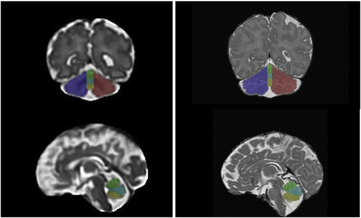
Using a prospective study design, high resolution anatomic magnetic resonance images (MRI) were acquired in PT infants (gestational age at birth < 32 weeks; birthweight < 1500 g) without cerebellar injury and healthy full-term controls. PT infants completed two MRIs, one as soon as medically stable and the other around TEA. Controls also completed two MRIs, one in utero (i.e. fetal MRI) and a postnatal MRI shortly after birth. The cerebellum of each participant was parcellated into 5 regions: left and right hemispheres, the anterior, neo and posterior vermis. Evidence of differences in regional volumes between term and pre-term infants matched for gestational age (GA) at the time of the first MRI were assessed using multiple linear regression.

RESULTS

WE STUDIED 76 SUBJECTS: 38 PT infants were matched to 38 healthy fetuses. At MRI-1, PT infants demonstrated decreased cerebellar hemispheric volumes and increased anterior, neo- and posterior vermian regional volumes when compared to healthy fetuses. At TEA, PT infants demonstrated a persistent increase in anterior, neo- and posterior vermian regional volumes but no longer showed reductions in cerebellar hemispheric volume. Only the neovermis volume demonstrated a significant negative association with birthweight, male gender and supratentorial injury.

CONCLUSIONS

In the absence of demonstrable cerebellar parenchymal injury evident on conventional MRI, PT birth is associated with cerebellar growth alterations that are regionally- and temporally-specific.

摘要

目的

比较极早产儿(PT)和健康胎龄匹配胎儿在 PT 期和胎龄相等期(TEA)的两个时间点的第三孕期小脑整体和区域体积生长。

研究设计

采用前瞻性研究设计,对无小脑损伤的 PT 婴儿(出生胎龄 < 32 周;出生体重 < 1500 克)和健康足月对照组进行高分辨率解剖磁共振成像(MRI)检查。PT 婴儿完成了两次 MRI,一次是在医学上稳定后,另一次是在 TEA 左右。对照组也完成了两次 MRI,一次是在宫内(即胎儿 MRI),一次是在出生后不久。每个参与者的小脑被分为 5 个区域:左半球和右半球、前叶、新叶和后叶。使用多元线性回归评估了与第一次 MRI 时胎龄(GA)相匹配的足月和早产儿之间区域体积差异的证据。

结果

我们研究了 76 名受试者:38 名 PT 婴儿与 38 名健康胎儿相匹配。在 MRI-1 时,与健康胎儿相比,PT 婴儿的小脑半球体积减小,前叶、新叶和后叶区域体积增大。在 TEA 时,PT 婴儿的前叶、新叶和后叶区域体积持续增加,但小脑半球体积不再减少。只有新叶体积与出生体重、男性性别和幕上损伤呈显著负相关。

结论

在没有明显的小脑实质损伤的情况下,PT 出生与小脑生长改变有关,这些改变具有区域性和时间特异性。

https://cdn.ncbi.nlm.nih.gov/pmc/blobs/20a7/6412008/6656bb005bec/gr1.jpg

相似文献

1
Early extra-uterine exposure alters regional cerebellar growth in infants born preterm.早期宫外暴露改变早产儿小脑区域生长。
Neuroimage Clin. 2019;21:101646. doi: 10.1016/j.nicl.2018.101646. Epub 2018 Dec 19.
2
Altered local cerebellar and brainstem development in preterm infants.早产儿小脑和脑干局部发育改变。
Neuroimage. 2020 Jun;213:116702. doi: 10.1016/j.neuroimage.2020.116702. Epub 2020 Mar 5.
3
Third Trimester Brain Growth in Preterm Infants Compared With In Utero Healthy Fetuses.与宫内健康胎儿相比,早产儿孕晚期的脑发育情况
Pediatrics. 2016 Nov;138(5). doi: 10.1542/peds.2016-1640.
4
Late gestation cerebellar growth is rapid and impeded by premature birth.妊娠晚期小脑生长迅速,且会因早产而受到阻碍。
Pediatrics. 2005 Mar;115(3):688-95. doi: 10.1542/peds.2004-1169.
5
Regional volumetric assessment of the brain in moderately preterm infants (30-35 gestational weeks) scanned at term-equivalent age on magnetic resonance imaging.在足月等效年龄时接受磁共振成像扫描的中度早产儿(孕龄30 - 35周)脑区容积评估
Early Hum Dev. 2017 Aug;111:36-41. doi: 10.1016/j.earlhumdev.2017.05.009. Epub 2017 May 30.
6
Changes in neonatal regional brain volume associated with preterm birth and perinatal factors.与早产及围生期因素相关的新生儿区域性脑容量变化。
Neuroimage. 2019 Jan 15;185:654-663. doi: 10.1016/j.neuroimage.2018.07.021. Epub 2018 Jul 21.
7
Brainstem and cerebellar volumes at magnetic resonance imaging are smaller in fetuses with congenital heart disease.磁共振成像显示,先天性心脏病胎儿的脑干和小脑体积较小。
Am J Obstet Gynecol. 2022 Aug;227(2):282.e1-282.e15. doi: 10.1016/j.ajog.2022.03.030. Epub 2022 Mar 16.
8
Third Trimester Cerebellar Metabolite Concentrations are Decreased in Very Premature Infants with Structural Brain Injury.晚期早产儿脑损伤与第三孕期小脑代谢物浓度降低相关。
Sci Rep. 2019 Feb 4;9(1):1212. doi: 10.1038/s41598-018-37203-4.
9
Longitudinal Preterm Cerebellar Volume: Perinatal and Neurodevelopmental Outcome Associations.纵向早产儿小脑体积:围产期和神经发育结局的关联。
Cerebellum. 2018 Oct;17(5):610-627. doi: 10.1007/s12311-018-0946-1.
10
Early life predictors of brain development at term-equivalent age in infants born across the gestational age spectrum.在跨越整个胎龄范围的足月出生婴儿中,预测其在胎龄相等时大脑发育的早期生命因素。
Neuroimage. 2019 Jan 15;185:813-824. doi: 10.1016/j.neuroimage.2018.04.031. Epub 2018 Apr 13.

引用本文的文献

1
Similarities and differences in cerebellar alterations between youth born preterm and youth born with congenital heart disease.早产青年与先天性心脏病青年小脑改变的异同
Sci Rep. 2025 Mar 26;15(1):10420. doi: 10.1038/s41598-025-94584-z.
2
The overgrowth of structure-function coupling in premature brain during infancy.婴儿期早产大脑中结构 - 功能耦合的过度生长。
Dev Cogn Neurosci. 2025 Apr;72:101535. doi: 10.1016/j.dcn.2025.101535. Epub 2025 Feb 21.
3
Cerebellar Development and the Burden of Prematurity.小脑发育与早产负担
Cerebellum. 2025 Jan 30;24(2):39. doi: 10.1007/s12311-025-01790-6.
4
Semi-automatic segmentation of the fetal brain from magnetic resonance imaging.基于磁共振成像的胎儿脑部半自动分割
Front Neurosci. 2022 Nov 11;16:1027084. doi: 10.3389/fnins.2022.1027084. eCollection 2022.
5
Systemic postnatal corticosteroids and magnetic resonance imaging measurements of corpus callosum and cerebellum of extremely preterm infants.系统产后皮质类固醇和极早产儿胼胝体和小脑的磁共振成像测量。
J Paediatr Child Health. 2023 Feb;59(2):282-287. doi: 10.1111/jpc.16286. Epub 2022 Nov 20.
6
Commentary - Cerebellar underdevelopment in the very preterm infant: Important and underestimated source of cognitive deficits.述评——极早产儿小脑发育不全:认知缺陷的重要且被低估的根源
J Neonatal Perinatal Med. 2021;14(4):451-456. doi: 10.3233/NPM-210774.
7
Developmental score of the infant brain: characterizing diffusion MRI in term- and preterm-born infants.婴儿大脑发育评分:足月和早产儿弥散磁共振成像特征。
Brain Struct Funct. 2020 Nov;225(8):2431-2445. doi: 10.1007/s00429-020-02132-4. Epub 2020 Aug 17.
8
Altered local cerebellar and brainstem development in preterm infants.早产儿小脑和脑干局部发育改变。
Neuroimage. 2020 Jun;213:116702. doi: 10.1016/j.neuroimage.2020.116702. Epub 2020 Mar 5.
9
Modelling brain development to detect white matter injury in term and preterm born neonates.建模大脑发育以检测足月和早产儿的脑白质损伤。
Brain. 2020 Feb 1;143(2):467-479. doi: 10.1093/brain/awz412.