Center for the Developing Brain, Children's National Hospital, Washington, D.C., USA.
Department of Psychology, American University, Washington, D.C., USA.
Neuroimage. 2020 Jun;213:116702. doi: 10.1016/j.neuroimage.2020.116702. Epub 2020 Mar 5.
Premature birth is associated with high prevalence of neurodevelopmental impairments in surviving infants. The putative role of cerebellar and brainstem dysfunction remains poorly understood, particularly in the absence of overt structural injury.
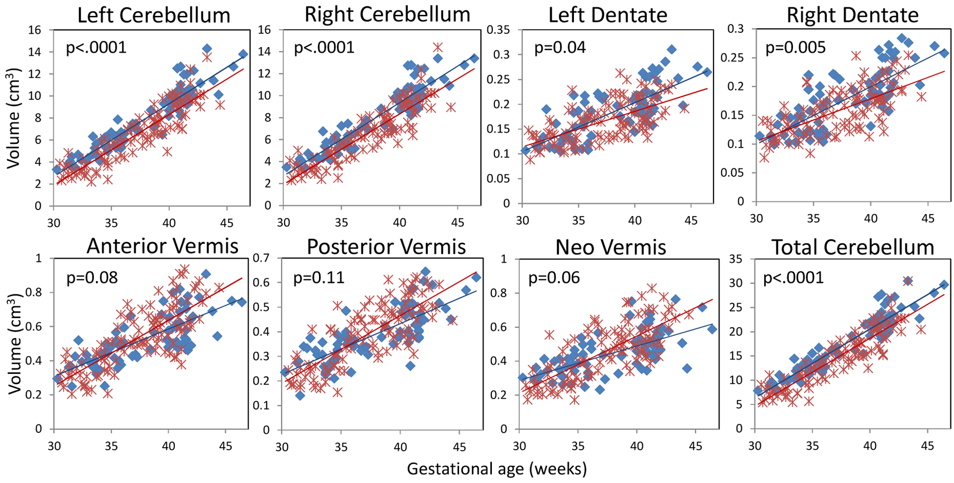
We compared in-utero versus ex-utero global, regional and local cerebellar and brainstem development in healthy fetuses (n = 38) and prematurely born infants without evidence of structural brain injury on conventional MRI studies (n = 74) that were performed at two time points: the first corresponding to the third trimester, either in utero or ex utero in the early postnatal period following preterm birth (30-40 weeks of gestation; 38 control fetuses; 52 premature infants) and the second at term equivalent age (37-46 weeks; 38 control infants; 58 premature infants). We compared 1) volumetric growth of 7 regions in the cerebellum (left and right hemispheres, left and right dentate nuclei, and the anterior, neo, and posterior vermis); 2) volumetric growth of 3 brainstem regions (midbrain, pons, and medulla); and 3) shape development in the cerebellum and brainstem using spherical harmonic description between the two groups.
Both premature and control groups showed regional cerebellar differences in growth rates, with the left and right cerebellar hemispheres showing faster growth compared to the vermis. In the brainstem, the pons grew faster than the midbrain and medulla in both prematurely born infants and controls. Using shape analyses, premature infants had smaller left and right cerebellar hemispheres but larger regional vermis and paravermis compared to in-utero control fetuses. For the brainstem, premature infants showed impaired growth of the superior surface of the midbrain, anterior surface of the pons, and inferior aspects of the medulla compared to the control fetuses. At term-equivalent age, premature infants had smaller cerebellar hemispheres bilaterally, extending to the superior aspect of the left cerebellar hemisphere, and larger anterior vermis and posteroinferior cerebellar lobes than healthy newborns. For the brainstem, large differences between premature infants and healthy newborns were found in the anterior surface of the pons.
This study analyzed both volumetric growth and shape development of the cerebellum and brainstem in premature infants compared to healthy fetuses using longitudinal MRI measurements. The findings in the present study suggested that preterm birth may alter global, regional and local development of the cerebellum and brainstem even in the absence of structural brain injury evident on conventional MRI.
早产儿出生后神经发育障碍的发生率较高。小脑和脑干功能障碍的潜在作用仍知之甚少,尤其是在没有明显结构损伤的情况下。
我们比较了健康胎儿(n=38)和无明显结构脑损伤的早产儿(n=74)在宫内和宫外的小脑和脑干的整体、区域和局部发育情况。这些早产儿是通过两次 MRI 研究获得的:第一次是在妊娠晚期(妊娠 30-40 周),在宫内或早产出生后的早期(胎龄 30-40 周);第二次是在相当于足月时(胎龄 37-46 周)。我们比较了 1)小脑 7 个区域(左、右半球,左、右齿状核和前、新、后蚓部)的体积生长;2)3 个脑干区域(中脑、脑桥和延髓)的体积生长;3)两组间小脑和脑干的形状发育。
早产组和对照组的小脑区域生长速度均存在差异,左、右小脑半球的生长速度快于蚓部。在脑干中,脑桥的生长速度快于中脑和延髓,无论是早产儿还是对照组都是如此。通过形态分析,与宫内对照组胎儿相比,早产儿的左、右小脑半球较小,但区域蚓部和旁蚓部较大。对于脑干,与对照组胎儿相比,早产儿的中脑上表面、脑桥前表面和延髓下表面的生长发育受损。在相当于足月时,早产儿的双侧小脑半球较小,左侧小脑半球上表面也较小,前蚓部和后下小脑叶较大。对于脑干,早产儿与正常新生儿之间存在较大差异。
本研究通过纵向 MRI 测量,分析了早产儿与健康胎儿的小脑和脑干的体积生长和形态发育。本研究的结果表明,即使在常规 MRI 上没有明显的结构性脑损伤,早产儿的小脑和脑干的整体、区域和局部发育可能会受到影响。