Yuan Yu-Song, Niu Su-Ping, Yu You-Lai, Zhang Pei-Xun, Yin Xiao-Feng, Han Na, Zhang Ya-Jun, Zhang Dian-Ying, Xu Hai-Lin, Kou Yu-Hui, Jiang Bao-Guo
Peking University People's Hospital, Beijing, China.
The Affiliated Hospital of Xuzhou Medical University, Xuzhou, Jiangsu Province, China.
Neural Regen Res. 2019 Apr;14(4):699-705. doi: 10.4103/1673-5374.247474.
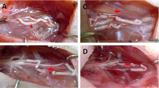
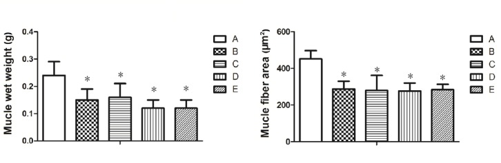
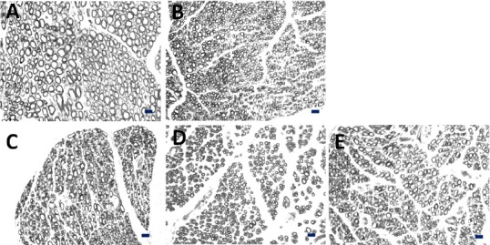
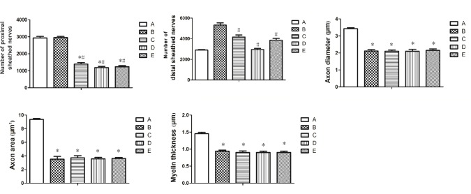
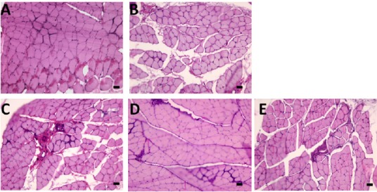
Our previous studies have confirmed that during nerve transposition repair to injured peripheral nerves, the regenerated nerve fibers of motor neurons in the anterior horn of the spinal cord can effectively repair distal nerve and target muscle tissue and restore muscle motor function. To observe the effect of nerve regeneration and motor function recovery after several types of nerve transposition for median nerve defect (2 mm), 30 Sprague-Dawley rats were randomly divided into sham operation group, epineurial neurorrhaphy group, musculocutaneous nerve transposition group, medial pectoral nerve transposition group, and radial nerve muscular branch transposition group. Three months after nerve repair, the wrist flexion test was used to evaluate the recovery of wrist flexion after regeneration of median nerve in the affected limbs of rats. The number of myelinated nerve fibers, the thickness of myelin sheath, the diameter of axons and the cross-sectional area of axons in the proximal and distal segments of the repaired nerves were measured by osmic acid staining. The ratio of newly produced distal myelinated nerve fibers to the number of proximal myelinated nerve fibers was calculated. Wet weights of the flexor digitorum superficialis muscles were measured. Muscle fiber morphology was detected using hematoxylin-eosin staining. The cross-sectional area of muscle fibers was calculated to assess the recovery of muscles. Results showed that wrist flexion function was restored, and the nerve grew into the distal effector in all three nerve transposition groups and the epineurial neurorrhaphy group. There were differences in the number of myelinated nerve fibers in each group. The magnification of proximal to distal nerves was 1.80, 3.00, 2.50, and 3.12 in epineurial neurorrhaphy group, musculocutaneous nerve transposition group, medial pectoral nerve transposition group, and radial nerve muscular branch transposition group, respectively. Nevertheless, axon diameters of new nerve fibers, cross-sectional areas of axons, thicknesses of myelin sheath, wet weights of flexor digitorum superficialis muscle and cross-sectional areas of muscle fibers of all three groups of donor nerves from different anterior horn motor neurons after nerve transposition were similar to those in the epineurial neurorrhaphy group. Our findings indicate that donor nerve translocation from different anterior horn motor neurons can effectively repair the target organs innervated by the median nerve. The corresponding spinal anterior horn motor neurons obtain functional reinnervation and achieve some degree of motor function in the affected limbs.
我们之前的研究已经证实,在对受损周围神经进行神经移位修复时,脊髓前角运动神经元再生的神经纤维能够有效修复远端神经和靶肌肉组织,并恢复肌肉运动功能。为了观察几种类型的神经移位对正中神经缺损(2毫米)后神经再生及运动功能恢复的影响,将30只Sprague-Dawley大鼠随机分为假手术组、神经外膜缝合组、肌皮神经移位组、胸内侧神经移位组和桡神经肌支移位组。神经修复3个月后,采用腕关节屈曲试验评估大鼠患侧肢体正中神经再生后腕关节屈曲的恢复情况。通过锇酸染色测量修复神经近端和远端节段有髓神经纤维数量、髓鞘厚度、轴突直径和轴突横截面积。计算新生远端有髓神经纤维数量与近端有髓神经纤维数量的比值。测量指浅屈肌湿重。采用苏木精-伊红染色检测肌纤维形态。计算肌纤维横截面积以评估肌肉恢复情况。结果显示,在所有三个神经移位组和神经外膜缝合组中,腕关节屈曲功能均得以恢复,且神经长入远端效应器。各组有髓神经纤维数量存在差异。神经外膜缝合组、肌皮神经移位组、胸内侧神经移位组和桡神经肌支移位组近端与远端神经的放大倍数分别为1.80、3.00、2.50和3.12。然而,神经移位后来自不同前角运动神经元的所有三组供体神经的新生神经纤维轴突直径、轴突横截面积、髓鞘厚度以及指浅屈肌湿重和肌纤维横截面积与神经外膜缝合组相似。我们的研究结果表明,来自不同前角运动神经元的供体神经移位能够有效修复正中神经支配的靶器官。相应的脊髓前角运动神经元获得功能性再支配,并在患侧肢体实现一定程度的运动功能。