Koga Naomichi, Kubo Nobuhide, Saeki Hiroshi, Sasaki Shun, Jogo Tomoko, Hirose Kosuke, Nakashima Yuichiro, Oki Eiji, Koga Yutaka, Oda Yoshinao, Oiwa Hisao, Oiwa Toshio, Maehara Yoshihiko
Department of Surgery and Science, Graduate School of Medical Sciences, Kyushu University, 3-1-1, Maidashi, Higashi-ku, Fukuoka, 812-8582, Japan.
Department of Anatomic Pathology, Pathological Sciences, Graduate School of Medical Sciences, Kyushu University, 3-1-1, Maidashi, Higashi-ku, Fukuoka, 812-8582, Japan.
Surg Case Rep. 2019 Jan 11;5(1):4. doi: 10.1186/s40792-019-0564-2.
Primary amelanotic malignant melanoma of esophagus, which is a subtype of primary malignant melanoma of the esophagus (PMME), is a very rare disease with a poor prognosis. We herein report a case of the amelanotic type of PMME.
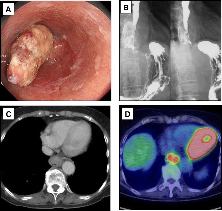
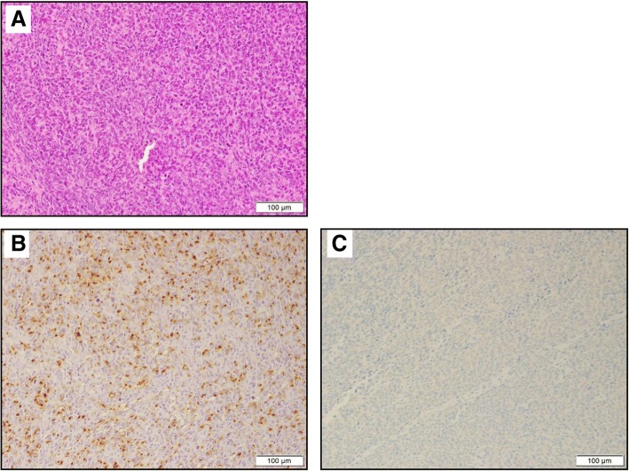
An 86-year-old woman was admitted to our hospital with symptoms of dysphagia. An endoscopic examination and constructed radiography revealed an elevated and semipedunculated lesion with an ulcer in the lower thoracic esophagus accompanied by another submucosal lesion of the esophagus. She was diagnosed with esophageal squamous cell carcinoma by a preoperative endoscopic biopsy. We performed thoracoscopy- and laparoscopy-assisted subtotal esophagectomy with lymphadenectomy. Based on the surgical specimens, although there were no melanocytes, we made a diagnosis of a malignant melanoma immunohistochemically; the tumor cells were positive for S-100 protein and HMB45 focally and partially for Melan-A.
We experienced a case of primary amelanotic malignant melanoma, and the patient has remained disease-free for 1 year since the surgery. Since the diagnosis of amelanotic type of PMME is difficult, it should be made by the combination of a morphological examination, pathological examination, and immunohistochemistry.
原发性食管无色素性恶性黑色素瘤是原发性食管恶性黑色素瘤(PMME)的一种亚型,是一种非常罕见且预后较差的疾病。我们在此报告一例无色素型PMME病例。
一名86岁女性因吞咽困难症状入院。内镜检查和造影显示胸段食管下段有一个隆起的半蒂状病变伴溃疡,同时食管还有另一个黏膜下病变。术前内镜活检诊断为食管鳞状细胞癌。我们进行了胸腔镜和腹腔镜辅助下食管次全切除术及淋巴结清扫术。基于手术标本,尽管未发现黑素细胞,但免疫组化诊断为恶性黑色素瘤;肿瘤细胞S-100蛋白和HMB45呈局灶阳性,Melan-A呈部分阳性。
我们遇到一例原发性无色素性恶性黑色素瘤病例,患者术后1年无疾病复发。由于无色素型PMME的诊断困难,应结合形态学检查、病理检查和免疫组化进行诊断。