Experimental and Clinical Pharmacopsychology, Department of Psychiatry, Psychotherapy, and Psychosomatics, Psychiatric Hospital, University of Zurich, Zurich, Switzerland.
Division Neuropsychology, Department of Psychology, University of Zurich, Zurich, Switzerland.
Neuroimage Clin. 2019;21:101652. doi: 10.1016/j.nicl.2019.101652. Epub 2019 Jan 4.
Cocaine use has been consistently associated with decreased gray matter volumes in the prefrontal cortex. However, it is unclear if such neuroanatomical abnormalities depict either pre-existing vulnerability markers or drug-induced consequences. Thus, this longitudinal MRI study investigated neuroplasticity and cognitive changes in relation to altered cocaine intake.
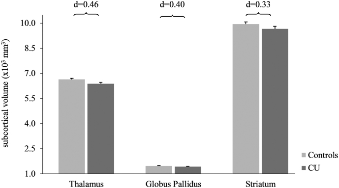
Surface-based morphometry, cocaine hair concentration, and cognitive performance were measured in 29 cocaine users (CU) and 38 matched controls at baseline and follow-up. Based on changes in hair cocaine concentration, CU were classified either as Decreasers (n = 15) or Sustained Users (n = 14). Surface-based morphometry measures did not include regional tissue volumes.
At baseline, CU displayed reduced cortical thickness (CT) in lateral frontal regions, and smaller cortical surface area (CSA) in the anterior cingulate cortex, compared to controls. In Decreasers, CT of the lateral frontal cortex increased whereas CT within the same regions tended to further decrease in Sustained Users. In contrast, no changes were found for CSA and subcortical structures. Changes in CT were linked to cognitive performance changes and amount of cocaine consumed over the study period.
These results suggest that frontal abnormalities in CU are partially drug-induced and can recover with decreased substance use. Moreover, recovery of frontal CT is accompanied by improved cognitive performance confirming that cognitive decline associated with cocaine use is potentially reversible.
可卡因的使用与前额叶皮质的灰质体积减少有关。然而,目前尚不清楚这些神经解剖异常是代表预先存在的脆弱性标志物还是药物引起的后果。因此,这项纵向 MRI 研究调查了与可卡因摄入量改变相关的神经可塑性和认知变化。
在基线和随访时,对 29 名可卡因使用者(CU)和 38 名匹配的对照者进行了基于表面的形态测量、头发可卡因浓度和认知表现的测量。根据头发可卡因浓度的变化,CU 被分为减少者(n=15)或持续使用者(n=14)。基于表面的形态测量值不包括区域组织体积。
在基线时,与对照组相比,CU 显示外侧额区皮质厚度(CT)降低,前扣带皮质皮质表面积(CSA)减小。在减少者中,外侧额叶皮质的 CT 增加,而在持续使用者中,同一区域的 CT 则有进一步降低的趋势。相比之下,CSA 和皮质下结构没有变化。CT 的变化与认知表现的变化和研究期间消耗的可卡因量有关。
这些结果表明,CU 中的额叶异常部分是药物诱导的,随着物质使用量的减少可以恢复。此外,额叶 CT 的恢复伴随着认知表现的改善,这证实了与可卡因使用相关的认知下降是可能可逆的。