Wong Thomas Y, Jesse Mary K, Jensen Alexandria, Kraeutler Matthew J, Coleman Christopher, Mei-Dan Omer
Department of Radiology, University of Colorado School of Medicine, Aurora, CO, USA.
Department of Musculoskeletal Radiology, University of Colorado School of Medicine, Aurora, CO, USA.
J Hip Preserv Surg. 2018 Dec 1;5(4):435-442. doi: 10.1093/jhps/hny042. eCollection 2018 Dec.
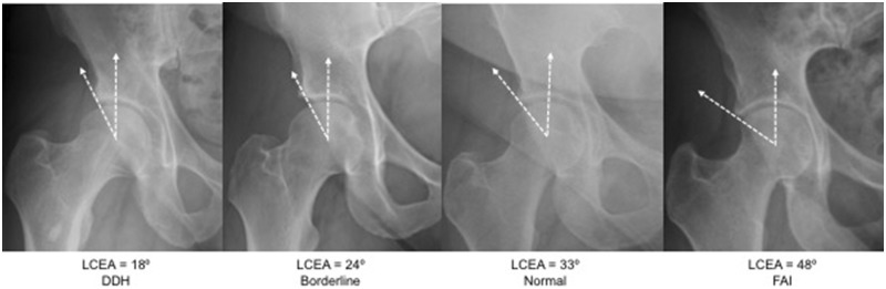
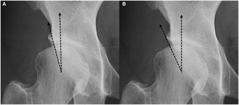
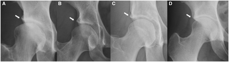
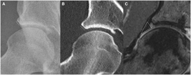
While radiographic findings of frank hip dysplasia are well defined, there is a lack of diagnostic criteria for patients with radiographically 'normal' hips who have borderline morphologic deficits and clinical instability. In this study, we aim to define and validate a new radiographic finding associated with hip instability known as the upsloping lateral sourcil (ULS). Patients (316) were reviewed for lateral center edge angles, generalized joint laxity assessed with the Beighton Hypermobility Score and the presence of the ULS. The ULS was defined as a caudal-to-cranial inclination of the middle-to-far lateral aspect of the acetabular sourcil with loss of the normal lateral acetabular concavity. The prevalence of the ULS correspondingly increased with the degree of under-coverage as defined by LCEA. Within the normal coverage group, hips with a ULS had smaller LCEAs than those without ULS (29° versus 32°, < 0.001). Among hips with a ULS, 59.00% had generalized joint laxity. The association between the ULS finding and generalized joint laxity was statistically significant ( < 0.01). The ULS is seen with higher prevalence in patients with clinical hip laxity and radiographically decreasing LCEA and may serve as an adjunctive finding in patients presenting with hip pain and instability. The ULS may help to characterize patients with borderline hip dysplasia and laxity that fall outside conventional imaging criteria for dysplasia.
虽然明显的髋关节发育不良的影像学表现已明确界定,但对于髋关节影像学“正常”但形态学存在临界缺陷且临床不稳定的患者,缺乏诊断标准。在本研究中,我们旨在定义并验证一种与髋关节不稳定相关的新影像学表现,即上斜外侧髋臼缘(ULS)。对316例患者的外侧中心边缘角、用贝顿关节活动过度评分评估的全身关节松弛情况以及ULS的存在情况进行了评估。ULS被定义为髋臼缘中外侧至远外侧从尾侧向颅侧的倾斜,同时正常外侧髋臼凹陷消失。ULS的患病率随外侧中心边缘角(LCEA)所定义的覆盖不足程度相应增加。在正常覆盖组中,有ULS的髋关节的LCEA小于无ULS的髋关节(29°对32°,P<0.001)。在有ULS的髋关节中,59.00%存在全身关节松弛。ULS表现与全身关节松弛之间的关联具有统计学意义(P<0.01)。在临床髋关节松弛且影像学上LCEA降低的患者中,ULS的患病率更高,并且在出现髋关节疼痛和不稳定的患者中可作为辅助表现。ULS可能有助于对处于髋关节发育不良和松弛临界状态且不符合传统发育不良影像学标准的患者进行特征描述。