Pathmanathan Angela U, McNair Helen A, Schmidt Maria A, Brand Douglas H, Delacroix Louise, Eccles Cynthia L, Gordon Alexandra, Herbert Trina, van As Nicholas J, Huddart Robert A, Tree Alison C
1 The Royal Marsden Hospital NHS Foundation Trust, Downs Road , Sutton , United Kingdom.
2 The Institute of Cancer Research, 15 Cotswold Road , Sutton , United Kingdom.
Br J Radiol. 2019 Mar;92(1095):20180948. doi: 10.1259/bjr.20180948. Epub 2019 Jan 24.
: With increasing incorporation of MRI in radiotherapy, we investigate two MRI sequences for prostate delineation in radiographer-led image guidance.
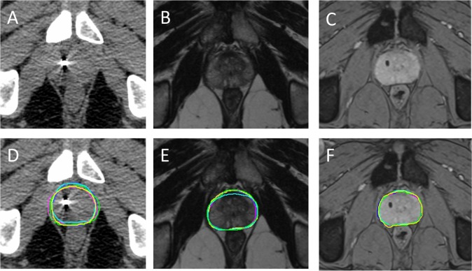
: Five therapeutic radiographers contoured the prostate individually on CT, T weighted (TW) and T* weighted (T*W) imaging for 10 patients. Contours were analysed with Monaco ADMIRE (research v. 2.0) to assess interobserver variability and accuracy by comparison with a gold standard clinician contour. Observers recorded time taken for contouring and scored image quality and confidence in contouring.
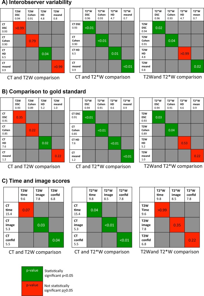
: There is good agreement when comparing radiographer contours to the gold-standard for all three imaging types with Dice similarity co-efficient 0.91-0.94, Cohen's κ 0.85-0.91, Hausdorff distance 4.6-7.6 mm and mean distance between contours 0.9-1.2 mm. In addition, there is good concordance between radiographers across all imaging modalities. Both TW and TW MRI show reduced interobserver variability and improved accuracy compared to CT, this was statistically significant for TW imaging compared to CT across all four comparison metrics. Comparing MRI sequences reveals significantly reduced interobserver variability and significantly improved accuracy on TW compared to TW MRI for DSC and Cohen's κ. Both MRI sequences scored significantly higher compared to CT for image quality and confidence in contouring, particularly TW. This was also reflected in the shorter time for contouring, measuring 15.4, 9.6 and 9.8 min for CT, TW and TW MRI respectively. Conclusion: Therapeutic radiographer prostate contours are more accurate, show less interobserver variability and are more confidently and quickly outlined on MRI compared to CT, particularly using TW MRI. Advances in knowledge: Our work is relevant for MRI sequence choice and development of the roles of the interprofessional team in the advancement of MRI-guided radiotherapy.
随着MRI在放射治疗中的应用日益增多,我们研究了两种MRI序列,用于在放射技师主导的图像引导下进行前列腺勾画。
五名放射治疗技师分别在CT、T加权(TW)和T加权(TW)成像上对10例患者的前列腺进行轮廓勾画。使用Monaco ADMIRE(研究版v.2.0)分析轮廓,通过与金标准临床医生轮廓进行比较,评估观察者间的变异性和准确性。观察者记录轮廓勾画所需时间,并对图像质量和轮廓勾画的信心进行评分。
将放射技师的轮廓与所有三种成像类型的金标准进行比较时,一致性良好,Dice相似系数为0.91 - 0.94,Cohen's κ为0.85 - 0.91,豪斯多夫距离为4.6 - 7.6毫米,轮廓间平均距离为0.9 - 1.2毫米。此外,所有成像模式下放射技师之间的一致性也很好。与CT相比,TW和TW MRI均显示观察者间变异性降低,准确性提高,在所有四个比较指标中,与CT相比,TW成像的差异具有统计学意义。比较MRI序列发现,与TW MRI相比,TW在DSC和Cohen's κ方面观察者间变异性显著降低,准确性显著提高。两种MRI序列在图像质量和轮廓勾画信心方面的评分均显著高于CT,尤其是TW。这也反映在轮廓勾画时间更短,CT、TW和TW MRI分别为15.4、9.6和9.8分钟。结论:与CT相比,放射治疗技师勾画的前列腺轮廓更准确,观察者间变异性更小,在MRI上勾画更有信心且速度更快,尤其是使用TW MRI。知识进展:我们的工作与MRI序列选择以及跨专业团队在MRI引导放射治疗进展中的角色发展相关。