• 文献检索
  • 文档翻译
  • 深度研究
  • 学术资讯
  • Suppr Zotero 插件Zotero 插件
  • 邀请有礼
  • 套餐&价格
  • 历史记录
应用&插件
Suppr Zotero 插件Zotero 插件浏览器插件Mac 客户端Windows 客户端微信小程序
定价
高级版会员购买积分包购买API积分包
服务
文献检索文档翻译深度研究API 文档MCP 服务
关于我们
关于 Suppr公司介绍联系我们用户协议隐私条款
关注我们

Suppr 超能文献

核心技术专利:CN118964589B侵权必究
粤ICP备2023148730 号-1Suppr @ 2026

文献检索

告别复杂PubMed语法,用中文像聊天一样搜索,搜遍4000万医学文献。AI智能推荐,让科研检索更轻松。

立即免费搜索

文件翻译

保留排版,准确专业,支持PDF/Word/PPT等文件格式,支持 12+语言互译。

免费翻译文档

深度研究

AI帮你快速写综述,25分钟生成高质量综述,智能提取关键信息,辅助科研写作。

立即免费体验

CNGA3 相关性无色素性视网膜炎的自适应光学视网膜成像:视网膜特征、双眼对称性和家族内变异性。

Adaptive Optics Retinal Imaging in CNGA3-Associated Achromatopsia: Retinal Characterization, Interocular Symmetry, and Intrafamilial Variability.

机构信息

UCL Institute of Ophthalmology, University College London, London, United Kingdom.

Moorfields Eye Hospital NHS Foundation Trust, City Road, London, United Kingdom.

出版信息

Invest Ophthalmol Vis Sci. 2019 Jan 2;60(1):383-396. doi: 10.1167/iovs.18-25880.

DOI:10.1167/iovs.18-25880
PMID:30682209
原文链接:https://pmc.ncbi.nlm.nih.gov/articles/PMC6354941/
Abstract

PURPOSE

To investigate retinal structure in subjects with CNGA3-associated achromatopsia and evaluate disease symmetry and intrafamilial variability.

METHODS

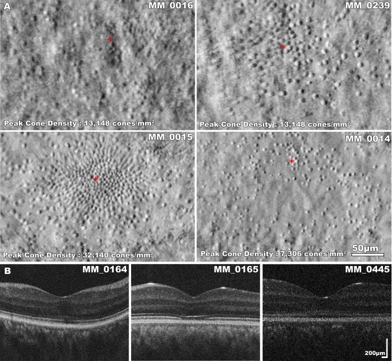
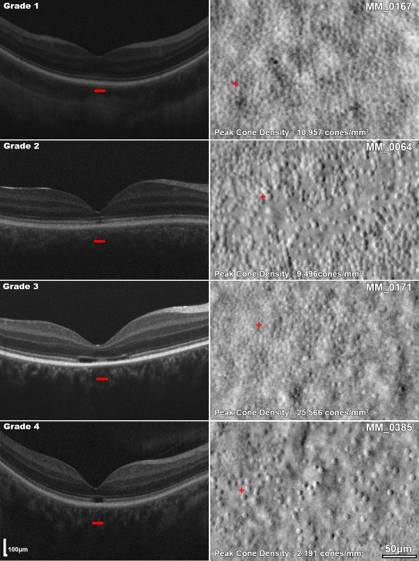
Thirty-eight molecularly confirmed subjects underwent ocular examination, optical coherence tomography (OCT), and nonconfocal split-detection adaptive optics scanning light ophthalmoscopy (AOSLO). OCT scans were used for evaluating foveal hypoplasia, grading foveal ellipsoid zone (EZ) disruption, and measuring outer nuclear layer (ONL) thickness. AOSLO images were used to quantify peak foveal cone density, intercell distance (ICD), and the coefficient of variation (CV) of ICD.

RESULTS

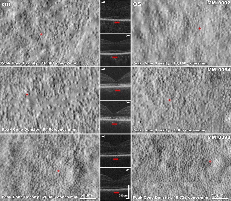
Mean (±SD) age was 25.9 (±13.1) years. Mean (± SD) best corrected visual acuity (BCVA) was 0.87 (±0.14) logarithm of the minimum angle of resolution. Examination with OCT showed variable disruption or loss of the EZ. Seven subjects were evaluated for disease symmetry, with peak foveal cone density, ICD, CV, ONL thickness, and BCVA not differing significantly between eyes. A cross-sectional evaluation of AOSLO imaging showed a mean (±SD) peak foveal cone density of 19,844 (±13,046) cones/mm2. There was a weak negative association between age and peak foveal cone density (r = -0.397, P = 0.102), as well as between EZ grade and age (P = 0.086).

CONCLUSIONS

The remnant cone mosaics were irregular and variably disrupted, with significantly lower peak foveal cone density than unaffected individuals. Variability was also seen among subjects with identical mutations. Therefore, subjects should be considered on an individual basis for stratification in clinical trials. Interocular symmetry suggests that both eyes have comparable therapeutic potential and the fellow eye can serve as a valid control. Longitudinal studies are needed, to further examine the weak negative association between age and foveal cone structure observed here.

摘要

目的

研究 CNGA3 相关色盲性视锥细胞营养不良患者的视网膜结构,评估疾病的对称性和家族内变异性。

方法

38 名经分子证实的患者接受了眼部检查、光学相干断层扫描(OCT)和非共焦分裂检测自适应光学扫描检眼镜(AOSLO)。OCT 扫描用于评估黄斑发育不良、黄斑椭圆区(EZ)分级破坏,并测量外核层(ONL)厚度。AOSLO 图像用于定量测量黄斑中心凹锥体细胞密度、细胞间距离(ICD)和 ICD 的变异系数(CV)。

结果

平均(±SD)年龄为 25.9(±13.1)岁。平均(±SD)最佳矫正视力(BCVA)为 0.87(±0.14)对数最小分辨角。OCT 检查显示 EZ 存在不同程度的破坏或缺失。7 名患者进行了疾病对称性评估,双眼黄斑中心凹锥体细胞密度、ICD、CV、ONL 厚度和 BCVA 无显著差异。AOSLO 成像的横断面评估显示平均(±SD)黄斑中心凹锥体细胞密度为 19844(±13046)个/毫米²。年龄与黄斑中心凹锥体细胞密度之间呈弱负相关(r=-0.397,P=0.102),EZ 分级与年龄之间也呈弱负相关(P=0.086)。

结论

残留的锥体细胞马赛克结构不规则且破坏程度不同,黄斑中心凹锥体细胞密度明显低于正常个体。具有相同突变的患者之间也存在差异。因此,应根据个体情况对临床试验中的患者进行分层。双眼的对称性表明双眼具有相当的治疗潜力,对侧眼可以作为有效的对照。需要进行纵向研究,以进一步探讨这里观察到的年龄与黄斑锥体细胞结构之间的弱负相关。

https://cdn.ncbi.nlm.nih.gov/pmc/blobs/b574/6354941/8eff53bc3552/i1552-5783-60-1-383-f05.jpg
https://cdn.ncbi.nlm.nih.gov/pmc/blobs/b574/6354941/098acdf2b598/i1552-5783-60-1-383-f01.jpg
https://cdn.ncbi.nlm.nih.gov/pmc/blobs/b574/6354941/8de1119f00b2/i1552-5783-60-1-383-f02.jpg
https://cdn.ncbi.nlm.nih.gov/pmc/blobs/b574/6354941/3b208bae11fc/i1552-5783-60-1-383-f03.jpg
https://cdn.ncbi.nlm.nih.gov/pmc/blobs/b574/6354941/715339f55f00/i1552-5783-60-1-383-f04.jpg
https://cdn.ncbi.nlm.nih.gov/pmc/blobs/b574/6354941/8eff53bc3552/i1552-5783-60-1-383-f05.jpg

相似文献

1
Adaptive Optics Retinal Imaging in CNGA3-Associated Achromatopsia: Retinal Characterization, Interocular Symmetry, and Intrafamilial Variability.CNGA3 相关性无色素性视网膜炎的自适应光学视网膜成像:视网膜特征、双眼对称性和家族内变异性。
Invest Ophthalmol Vis Sci. 2019 Jan 2;60(1):383-396. doi: 10.1167/iovs.18-25880.
2
Longitudinal Imaging of the Foveal Cone Mosaic in CNGA3-Associated Achromatopsia.CNGA3 相关性无色素性视锥营养不良的中心凹视锥细胞嵌合体的纵向成像。
Invest Ophthalmol Vis Sci. 2024 Oct 1;65(12):6. doi: 10.1167/iovs.65.12.6.
3
Characterization of Retinal Structure in ATF6-Associated Achromatopsia.ATF6 相关先天性静止性夜盲症的视网膜结构特征。
Invest Ophthalmol Vis Sci. 2019 Jun 3;60(7):2631-2640. doi: 10.1167/iovs.19-27047.
4
Interocular Symmetry of Foveal Cone Topography in Congenital Achromatopsia.先天性色觉缺失患者的中心凹锥细胞形态的双眼对称性。
Curr Eye Res. 2020 Oct;45(10):1257-1264. doi: 10.1080/02713683.2020.1737138. Epub 2020 Mar 13.
5
Residual Foveal Cone Structure in CNGB3-Associated Achromatopsia.CNGB3相关全色盲中的残余中央凹视锥结构
Invest Ophthalmol Vis Sci. 2016 Aug 1;57(10):3984-95. doi: 10.1167/iovs.16-19313.
6
Retinal structure and function in achromatopsia: implications for gene therapy.色盲症的视网膜结构和功能:对基因治疗的启示。
Ophthalmology. 2014 Jan;121(1):234-245. doi: 10.1016/j.ophtha.2013.08.017. Epub 2013 Oct 20.
7
REPEATABILITY AND LONGITUDINAL ASSESSMENT OF FOVEAL CONE STRUCTURE IN CNGB3-ASSOCIATED ACHROMATOPSIA.CNGB3相关全色盲中黄斑锥体细胞结构的可重复性及纵向评估
Retina. 2017 Oct;37(10):1956-1966. doi: 10.1097/IAE.0000000000001434.
8
Photoreceptor Structure in GNAT2-Associated Achromatopsia.GNAT2 相关先天性静止性夜盲症的光感受器结构。
Invest Ophthalmol Vis Sci. 2020 Mar 9;61(3):40. doi: 10.1167/iovs.61.3.40.
9
Deep Phenotyping of PDE6C-Associated Achromatopsia.PDE6C 相关先天性静止性夜盲症的深度表型分析。
Invest Ophthalmol Vis Sci. 2019 Dec 2;60(15):5112-5123. doi: 10.1167/iovs.19-27761.
10
The effect of cone opsin mutations on retinal structure and the integrity of the photoreceptor mosaic.视锥蛋白突变对视网膜结构和光感受器嵌合体完整性的影响。
Invest Ophthalmol Vis Sci. 2012 Dec 5;53(13):8006-15. doi: 10.1167/iovs.12-11087.

引用本文的文献

1
Telecentric model eye for correction of image distortion in adaptive optics ophthalmoscopes.用于自适应光学检眼镜图像畸变校正的远心模型眼。
Biomed Opt Express. 2025 Jun 16;16(7):2767-2791. doi: 10.1364/BOE.565589. eCollection 2025 Jul 1.
2
Longitudinal Imaging of the Parafoveal Cone Mosaic in Congenital Achromatopsia.先天性全色盲患者旁中央凹视锥细胞镶嵌的纵向成像
Ophthalmol Sci. 2025 Mar 14;5(4):100765. doi: 10.1016/j.xops.2025.100765. eCollection 2025 Jul-Aug.
3
Enhanced genotype-phenotype analysis using multimodal adaptive optics and 3D protein structure in Bietti Crystalline Dystrophy.

本文引用的文献

1
Assessing the Interocular Symmetry of Foveal Outer Nuclear Layer Thickness in Achromatopsia.评估全色盲患者中央凹外核层厚度的双眼对称性。
Transl Vis Sci Technol. 2019 Oct 2;8(5):21. doi: 10.1167/tvst.8.5.21. eCollection 2019 Sep.
2
Fast adaptive optics scanning light ophthalmoscope retinal montaging.快速自适应光学扫描激光检眼镜视网膜图像拼接
Biomed Opt Express. 2018 Aug 15;9(9):4317-4328. doi: 10.1364/BOE.9.004317. eCollection 2018 Sep 1.
3
Longitudinal Assessment of Retinal Structure in Achromatopsia Patients With Long-Term Follow-up.
在比埃蒂结晶状营养不良中使用多模态自适应光学和3D蛋白质结构进行增强的基因型-表型分析。
Am J Ophthalmol Case Rep. 2025 Mar 22;38:102312. doi: 10.1016/j.ajoc.2025.102312. eCollection 2025 Jun.
4
From Cellular to Metabolic: Advances in Imaging of Inherited Retinal Diseases.从细胞层面到代谢层面:遗传性视网膜疾病成像技术的进展
Diagnostics (Basel). 2024 Dec 26;15(1):28. doi: 10.3390/diagnostics15010028.
5
Longitudinal Imaging of the Foveal Cone Mosaic in CNGA3-Associated Achromatopsia.CNGA3 相关性无色素性视锥营养不良的中心凹视锥细胞嵌合体的纵向成像。
Invest Ophthalmol Vis Sci. 2024 Oct 1;65(12):6. doi: 10.1167/iovs.65.12.6.
6
Gene Therapy for Achromatopsia.基因治疗色盲症。
Int J Mol Sci. 2024 Sep 9;25(17):9739. doi: 10.3390/ijms25179739.
7
Multimodal in-vivo maps as a tool to characterize retinal structural biomarkers for progression in adult-onset Stargardt disease.多模态体内图谱作为一种工具,用于表征成人型Stargardt病进展中的视网膜结构生物标志物。
Front Ophthalmol (Lausanne). 2024 Apr 23;4:1384473. doi: 10.3389/fopht.2024.1384473. eCollection 2024.
8
Congenital Stationary Night Blindness: Structure, Function and Genotype-Phenotype Correlations in a Cohort of 122 Patients.先天性静止性夜盲症:122 例患者队列的结构、功能及基因型-表型相关性研究。
Ophthalmol Retina. 2024 Sep;8(9):932-941. doi: 10.1016/j.oret.2024.03.017. Epub 2024 Mar 24.
9
Cone Density Distribution and Related Factors in Patients Receiving Hydroxychloroquine Treatment.接受羟氯喹治疗的患者的锥体密度分布及相关因素。
Invest Ophthalmol Vis Sci. 2023 Sep 1;64(12):29. doi: 10.1167/iovs.64.12.29.
10
Cone Photoreceptor Morphology in Choroideremia Assessed Using Non-Confocal Split-Detection Adaptive Optics Scanning Light Ophthalmoscopy.应用非共焦分裂探测自适应光学扫描检眼镜评估色素性视网膜炎中的锥形光感受器形态。
Invest Ophthalmol Vis Sci. 2023 Jul 3;64(10):36. doi: 10.1167/iovs.64.10.36.
长时程随访先天性静止性夜盲症患者视网膜结构的纵向评估。
Invest Ophthalmol Vis Sci. 2018 Dec 3;59(15):5735-5744. doi: 10.1167/iovs.18-25452.
4
The Henle Fiber Layer in Albinism: Comparison to Normal and Relationship to Outer Nuclear Layer Thickness and Foveal Cone Density.白化病中的 Henle 纤维层:与正常的比较以及与外核层厚度和中心凹锥密度的关系。
Invest Ophthalmol Vis Sci. 2018 Nov 1;59(13):5336-5348. doi: 10.1167/iovs.18-24145.
5
Deep learning based detection of cone photoreceptors with multimodal adaptive optics scanning light ophthalmoscope images of achromatopsia.基于深度学习利用全色盲多模态自适应光学扫描激光检眼镜图像检测视锥光感受器
Biomed Opt Express. 2018 Jul 18;9(8):3740-3756. doi: 10.1364/BOE.9.003740. eCollection 2018 Aug 1.
6
Automatic Cone Photoreceptor Localisation in Healthy and Stargardt Afflicted Retinas Using Deep Learning.使用深度学习自动定位健康和斯特格德特视网膜中的锥形光感受器。
Sci Rep. 2018 May 21;8(1):7911. doi: 10.1038/s41598-018-26350-3.
7
Ultra-high contrast retinal display system for single photoreceptor psychophysics.用于单光感受器心理物理学的超高对比度视网膜显示系统。
Biomed Opt Express. 2017 Dec 8;9(1):157-172. doi: 10.1364/BOE.9.000157. eCollection 2018 Jan 1.
8
Achromatopsia: clinical features, molecular genetics, animal models and therapeutic options.色盲症:临床特征、分子遗传学、动物模型及治疗选择
Ophthalmic Genet. 2018 Apr;39(2):149-157. doi: 10.1080/13816810.2017.1418389. Epub 2018 Jan 5.
9
Adaptive optics imaging of inherited retinal diseases.遗传性视网膜疾病的自适应光学成像。
Br J Ophthalmol. 2018 Aug;102(8):1028-1035. doi: 10.1136/bjophthalmol-2017-311328. Epub 2017 Nov 15.
10
CNGB3 mutations cause severe rod dysfunction.CNGB3基因突变会导致严重的视杆细胞功能障碍。
Ophthalmic Genet. 2018 Jan-Feb;39(1):108-114. doi: 10.1080/13816810.2017.1368087. Epub 2017 Sep 20.