Department of Bioengineering, Imperial College London, South Kensington, SW7 2AZ London, United Kingdom.
Neurophysiology, Institute of Zoology, Universität Regensburg, 93040 Regensburg, Germany.
PLoS One. 2019 Jan 28;14(1):e0210564. doi: 10.1371/journal.pone.0210564. eCollection 2019.
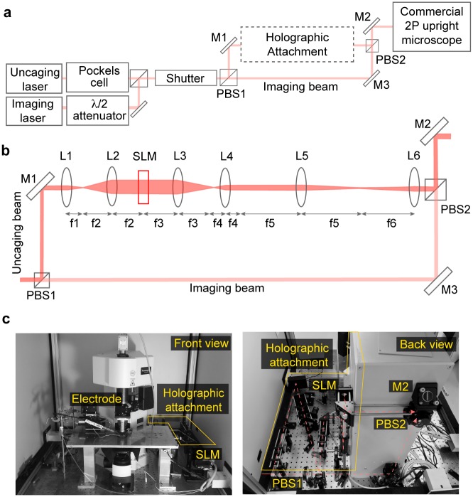
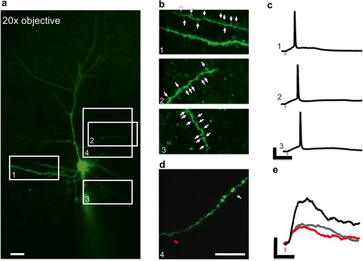
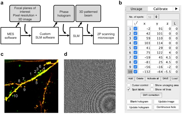
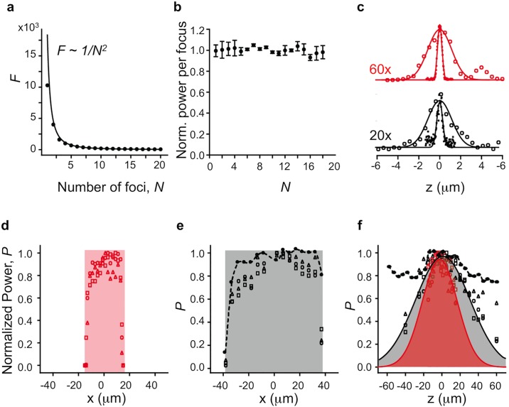
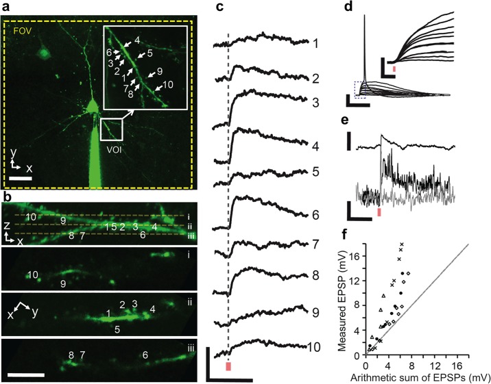
Patterned two-photon (2P) photolysis via holographic illumination is a powerful method to investigate neuronal function because of its capability to emulate multiple synaptic inputs in three dimensions (3D) simultaneously. However, like any optical system, holographic projectors have a finite space-bandwidth product that restricts the spatial range of patterned illumination or field-of-view (FOV) for a desired resolution. Such trade-off between holographic FOV and resolution restricts the coverage within a limited domain of the neuron's dendritic tree to perform highly resolved patterned 2P photolysis on individual spines. Here, we integrate a holographic projector into a commercial 2P galvanometer-based 2D scanning microscope with an uncaging unit and extend the accessible holographic FOV by using the galvanometer scanning mirrors to reposition the holographic FOV arbitrarily across the imaging FOV. The projector system utilizes the microscope's built-in imaging functions. Stimulation positions can be selected from within an acquired 3D image stack (the volume-of-interest, VOI) and the holographic projector then generates 3D illumination patterns with multiple uncaging foci. The imaging FOV of our system is 800×800 μm2 within which a holographic VOI of 70×70×70 μm3 can be chosen at arbitrary positions and also moved during experiments without moving the sample. We describe the design and alignment protocol as well as the custom software plugin that controls the 3D positioning of stimulation sites. We demonstrate the neurobiological application of the system by simultaneously uncaging glutamate at multiple spines within dendritic domains and consequently observing summation of postsynaptic potentials at the soma, eventually resulting in action potentials. At the same time, it is possible to perform two-photon Ca2+ imaging in 2D in the dendrite and thus to monitor synaptic Ca2+ entry in selected spines and also local regenerative events such as dendritic action potentials.
基于全息照明的图案化双光子(2P)光解是一种强大的研究神经元功能的方法,因为它能够在三维(3D)空间中同时模拟多个突触输入。然而,与任何光学系统一样,全息投影仪的空间带宽积有限,这限制了图案化照明的空间范围或所需分辨率的视野(FOV)。这种全息 FOV 和分辨率之间的权衡限制了在神经元树突的有限区域内的覆盖范围,以对单个棘突进行高分辨率的图案化 2P 光解。在这里,我们将全息投影仪集成到具有离笼单元的商用 2P 振镜式 2D 扫描显微镜中,并使用振镜扫描镜重新定位全息 FOV,从而在成像 FOV 内任意位置扩展可访问的全息 FOV。投影仪系统利用显微镜的内置成像功能。刺激位置可以从采集的 3D 图像堆栈(感兴趣体积,VOI)中选择,然后全息投影仪生成具有多个离笼焦点的 3D 照明图案。我们系统的成像 FOV 为 800×800 μm2,可以在任意位置选择 70×70×70 μm3 的全息 VOI,并且在实验过程中无需移动样品即可移动。我们描述了设计和对准协议以及控制刺激部位 3D 定位的定制软件插件。我们通过同时在树突域内的多个棘突处离笼谷氨酸来演示该系统的神经生物学应用,从而观察到胞体处的突触后电位总和,最终导致动作电位。同时,可以在树突中进行 2D 的双光子 Ca2+成像,从而监测选定棘突中的突触 Ca2+内流以及局部再生事件,如树突动作电位。