• 文献检索
  • 文档翻译
  • 深度研究
  • 学术资讯
  • Suppr Zotero 插件Zotero 插件
  • 邀请有礼
  • 套餐&价格
  • 历史记录
应用&插件
Suppr Zotero 插件Zotero 插件浏览器插件Mac 客户端Windows 客户端微信小程序
定价
高级版会员购买积分包购买API积分包
服务
文献检索文档翻译深度研究API 文档MCP 服务
关于我们
关于 Suppr公司介绍联系我们用户协议隐私条款
关注我们

Suppr 超能文献

核心技术专利:CN118964589B侵权必究
粤ICP备2023148730 号-1Suppr @ 2026

文献检索

告别复杂PubMed语法,用中文像聊天一样搜索,搜遍4000万医学文献。AI智能推荐,让科研检索更轻松。

立即免费搜索

文件翻译

保留排版,准确专业,支持PDF/Word/PPT等文件格式,支持 12+语言互译。

免费翻译文档

深度研究

AI帮你快速写综述,25分钟生成高质量综述,智能提取关键信息,辅助科研写作。

立即免费体验

甲状腺超声弹性成像:原理与现状

Ultrasound elastography of the thyroid: principles and current status.

作者信息

Zhao Chong-Ke, Xu Hui-Xiong

机构信息

Department of Medical Ultrasound, Shanghai Tenth People's Hospital, Ultrasound Research and Education Institute, Tongji University School of Medicine, Shanghai, China.

Thyroid Institute, Tongji University School of Medicine, Shanghai, China.

出版信息

Ultrasonography. 2019 Apr;38(2):106-124. doi: 10.14366/usg.18037. Epub 2018 Oct 1.

DOI:10.14366/usg.18037
PMID:30690960
原文链接:https://pmc.ncbi.nlm.nih.gov/articles/PMC6443591/
Abstract

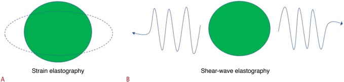
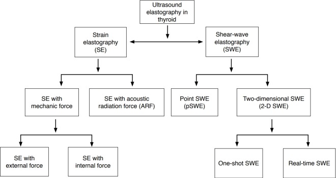
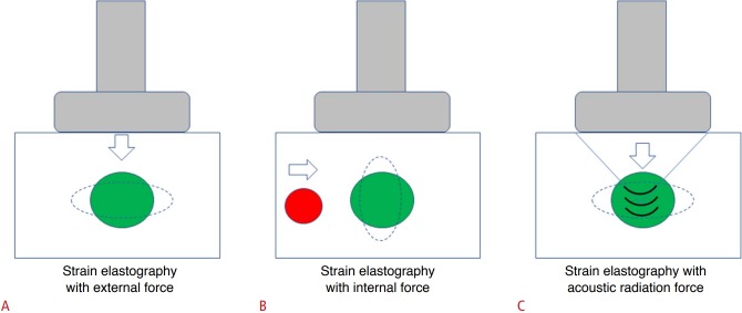
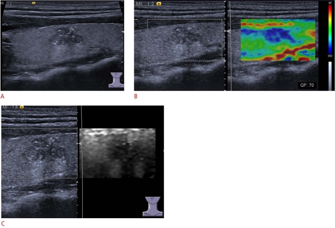
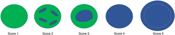
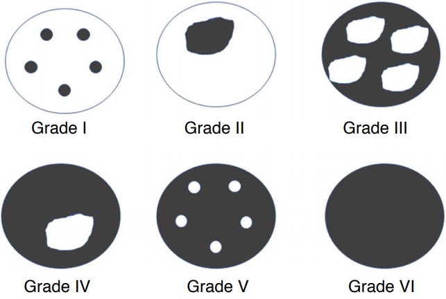
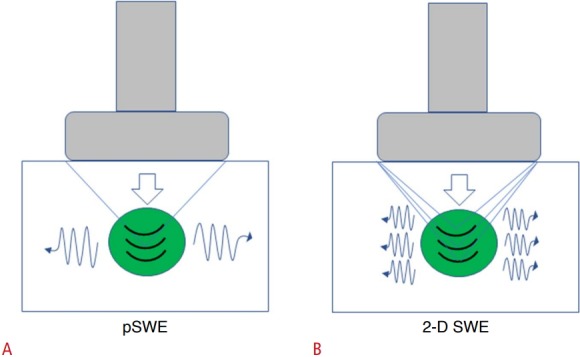
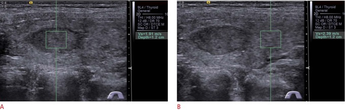
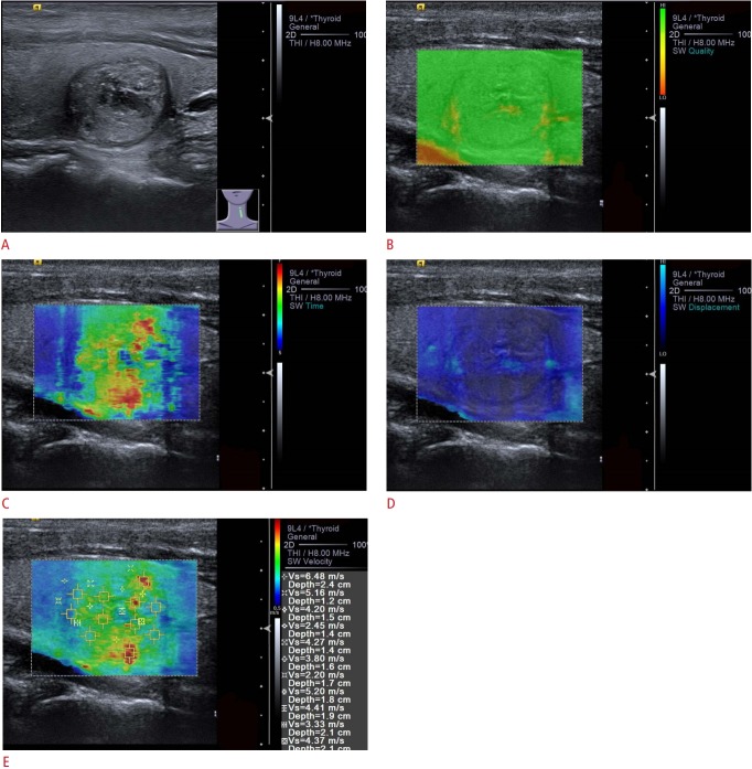
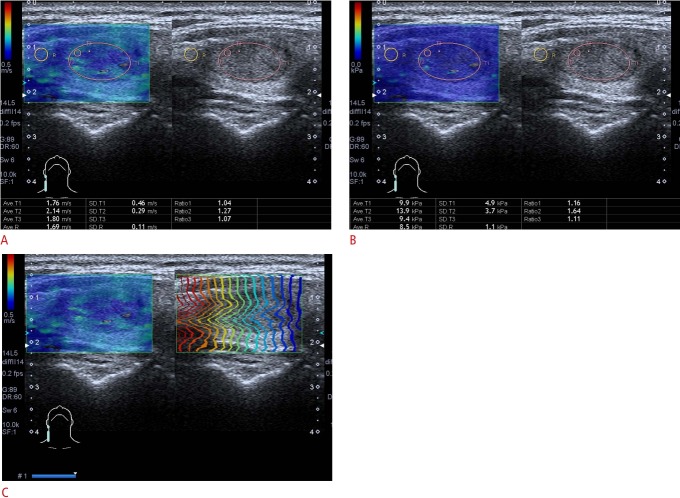
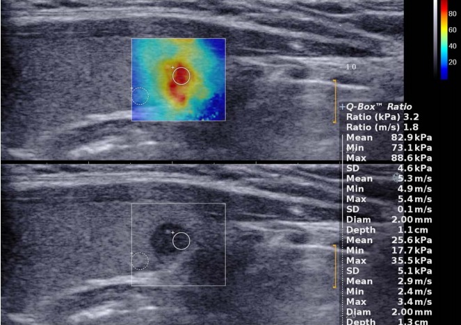
Ultrasound (US) elastography has been introduced as a non-invasive technique for evaluating thyroid diseases. This paper presents a detailed description of the technical principles, peculiarities, and limitations of US elastography techniques, including strain elastography and shear-wave elastography. This review was conducted from a clinical perspective, and aimed to assess the usefulness of US elastography for thyroid diseases in specific clinical scenarios. Although its main focus is on thyroid nodules, the applications of US elastography for other thyroid diseases, such as diffuse thyroid diseases and thyroiditis, are also presented. Furthermore, unresolved questions and directions for future research are also discussed.

摘要

超声(US)弹性成像已作为一种评估甲状腺疾病的非侵入性技术被引入。本文详细描述了超声弹性成像技术的技术原理、特点和局限性,包括应变弹性成像和剪切波弹性成像。本综述是从临床角度进行的,旨在评估超声弹性成像在特定临床场景中对甲状腺疾病的实用性。尽管其主要重点是甲状腺结节,但也介绍了超声弹性成像在其他甲状腺疾病,如弥漫性甲状腺疾病和甲状腺炎中的应用。此外,还讨论了未解决的问题和未来研究的方向。

https://cdn.ncbi.nlm.nih.gov/pmc/blobs/9017/6443591/3455c8e32545/usg-18037f13.jpg
https://cdn.ncbi.nlm.nih.gov/pmc/blobs/9017/6443591/f4a5e43872d5/usg-18037f1.jpg
https://cdn.ncbi.nlm.nih.gov/pmc/blobs/9017/6443591/ff11e3c8dd78/usg-18037f2.jpg
https://cdn.ncbi.nlm.nih.gov/pmc/blobs/9017/6443591/ed81f3100380/usg-18037f3.jpg
https://cdn.ncbi.nlm.nih.gov/pmc/blobs/9017/6443591/ba75de972fa3/usg-18037f4.jpg
https://cdn.ncbi.nlm.nih.gov/pmc/blobs/9017/6443591/7cf194d623af/usg-18037f5.jpg
https://cdn.ncbi.nlm.nih.gov/pmc/blobs/9017/6443591/1b82ec3f61d3/usg-18037f6.jpg
https://cdn.ncbi.nlm.nih.gov/pmc/blobs/9017/6443591/294238fcd431/usg-18037f7.jpg
https://cdn.ncbi.nlm.nih.gov/pmc/blobs/9017/6443591/5608ed5d46c0/usg-18037f8.jpg
https://cdn.ncbi.nlm.nih.gov/pmc/blobs/9017/6443591/29d46353ab7b/usg-18037f9.jpg
https://cdn.ncbi.nlm.nih.gov/pmc/blobs/9017/6443591/eccbd980bcc9/usg-18037f10.jpg
https://cdn.ncbi.nlm.nih.gov/pmc/blobs/9017/6443591/755afef97d8f/usg-18037f11.jpg
https://cdn.ncbi.nlm.nih.gov/pmc/blobs/9017/6443591/81e0f1bf514a/usg-18037f12.jpg
https://cdn.ncbi.nlm.nih.gov/pmc/blobs/9017/6443591/3455c8e32545/usg-18037f13.jpg

相似文献

1
Ultrasound elastography of the thyroid: principles and current status.甲状腺超声弹性成像:原理与现状
Ultrasonography. 2019 Apr;38(2):106-124. doi: 10.14366/usg.18037. Epub 2018 Oct 1.
2
Ultrasound elastography in thyroid disease.甲状腺疾病中的超声弹性成像
Med Ultrason. 2015 Mar;17(1):74-96. doi: 10.11152/mu.2013.2066.171.smd.
3
Guidelines and recommendations on the clinical use of shear wave elastography for evaluating thyroid nodule1.《甲状腺结节剪切波弹性成像临床应用指南及专家共识》1。
Clin Hemorheol Microcirc. 2019;72(1):39-60. doi: 10.3233/CH-180452.
4
Precision Medicine with 3D Ultrasound.三维超声精准医疗
VideoEndocrinology. 2020 Sep 7;7(3). doi: 10.1089/ve.2020.0191. eCollection 2020.
5
Quantitative assessment of thyroid gland elasticity with shear-wave elastography in pediatric patients with Hashimoto's thyroiditis.利用剪切波弹性成像技术对桥本甲状腺炎患儿甲状腺弹性进行定量评估。
J Med Ultrason (2001). 2018 Jul;45(3):417-423. doi: 10.1007/s10396-018-0859-0. Epub 2018 Jan 18.
6
Ultrasound elastography in the evaluation of thyroid pathology. Current status.超声弹性成像在甲状腺病变评估中的应用。现状。
Eur J Radiol. 2014 Mar;83(3):420-8. doi: 10.1016/j.ejrad.2013.05.008. Epub 2013 Jun 12.
7
Application of Various Additional Imaging Techniques for Thyroid Ultrasound: Direct Comparison of Combined Various Elastography and Doppler Parameters to Gray-Scale Ultrasound in Differential Diagnosis of Thyroid Nodules.多种甲状腺超声附加成像技术的应用:在甲状腺结节鉴别诊断中联合多种弹性成像和多普勒参数与灰阶超声的直接比较
Ultrasound Med Biol. 2018 Aug;44(8):1679-1686. doi: 10.1016/j.ultrasmedbio.2018.04.006. Epub 2018 May 22.
8
Shear wave elastography in the evaluation of level VI lymph nodes in papillary thyroid carcinoma: combined with gray-scale ultrasound ex vivo.剪切波弹性成像在评估甲状腺乳头状癌 VI 级淋巴结中的应用:结合灰阶超声的离体研究。
BMC Cancer. 2018 Oct 20;18(1):1001. doi: 10.1186/s12885-018-4897-1.
9
Diagnosis of Thyroid Nodule with New Ultrasound Imaging Modalities.利用新型超声成像模式诊断甲状腺结节
VideoEndocrinology. 2020 Mar 30;7(1). doi: 10.1089/ve.2020.0173. eCollection 2019.
10
Ultrasound elastography for thyroid nodules: recent advances.甲状腺结节的超声弹性成像:最新进展。
Ultrasonography. 2014 Apr;33(2):75-82. doi: 10.14366/usg.13025. Epub 2014 Feb 26.

引用本文的文献

1
Deep learning based motion correction in ultrasound microvessel imaging approach improves thyroid nodule classification.基于深度学习的超声微血管成像方法中的运动校正可改善甲状腺结节分类。
Sci Rep. 2025 May 30;15(1):19081. doi: 10.1038/s41598-025-02728-y.
2
Unusual nodular goiter with recurrent laryngeal nerve palsy due to severe degeneration caused by intense chronic inflammation: a case report with histopathological evidence and review of the literature.因严重慢性炎症导致严重变性而伴有喉返神经麻痹的罕见结节性甲状腺肿:一例有组织病理学证据的病例报告及文献复习
BMC Endocr Disord. 2025 May 12;25(1):128. doi: 10.1186/s12902-025-01929-w.
3

本文引用的文献

1
The value of shear wave elastography in diffuse thyroid disease.剪切波弹性成像在弥漫性甲状腺疾病中的价值。
Clin Imaging. 2018 May-Jun;49:187-192. doi: 10.1016/j.clinimag.2018.03.019. Epub 2018 Mar 29.
2
Differentiation of Benign and Malignant Thyroid Nodules by Using Comb-push Ultrasound Shear Elastography: A Preliminary Two-plane View Study.应用梳状推扫超声剪切弹性成像技术鉴别甲状腺良恶性结节:初步的双平面研究。
Acad Radiol. 2018 Nov;25(11):1388-1397. doi: 10.1016/j.acra.2018.02.025. Epub 2018 Mar 21.
3
Artifacts in Shear Wave Elastography Images of Thyroid Nodules.
Integrating shear wave elastography into clinical prediction of Graves' disease recurrence: a novel risk scoring system.
将剪切波弹性成像纳入Graves病复发的临床预测:一种新型风险评分系统。
Front Endocrinol (Lausanne). 2025 Mar 12;16:1551983. doi: 10.3389/fendo.2025.1551983. eCollection 2025.
4
Shear wave elastography for thyroid nodule evaluation in patients with chronic autoimmune thyroiditis.剪切波弹性成像在慢性自身免疫性甲状腺炎患者甲状腺结节评估中的应用
Endocrine. 2025 May;88(2):482-490. doi: 10.1007/s12020-025-04159-1. Epub 2025 Jan 19.
5
Management Aspects of Medical Therapy in Graves Disease.格雷夫斯病的药物治疗管理方面
Endocr Pract. 2025 Apr;31(4):536-546. doi: 10.1016/j.eprac.2024.12.012. Epub 2024 Dec 17.
6
Application of shear wave elastography in the management of thyroid nodules in children and adolescents: our experience and a review of the literature.剪切波弹性成像在儿童及青少年甲状腺结节管理中的应用:我们的经验及文献综述
Front Endocrinol (Lausanne). 2024 Nov 20;15:1486285. doi: 10.3389/fendo.2024.1486285. eCollection 2024.
7
Diagnostic Accuracy of Ultrasound Elastography Versus Fine-Needle Aspiration Cytology in Predicting Benign and Malignant Thyroid Nodules in Solitary and Multinodular Thyroid Glands: A Comparative Analysis.超声弹性成像与细针穿刺细胞学检查在预测单发性和多发性甲状腺结节的良恶性方面的诊断准确性:一项对比分析
Cureus. 2024 Nov 1;16(11):e72811. doi: 10.7759/cureus.72811. eCollection 2024 Nov.
8
Role of Ultrasound Elastography in Evaluating Suspicious Thyroid Nodules.超声弹性成像在评估可疑甲状腺结节中的作用
Indian J Surg Oncol. 2024 Dec;15(4):646-651. doi: 10.1007/s13193-024-01956-4. Epub 2024 May 21.
9
The Horizon of Thyroid Imaging Reporting and Data System in the Diagnostic Performance of Thyroid Nodules: Clinical Application and Future Perspectives.甲状腺影像报告和数据系统在甲状腺结节诊断性能中的应用前景:临床应用与未来展望
touchREV Endocrinol. 2024 Oct;20(2):81-90. doi: 10.17925/EE.2024.20.2.11. Epub 2024 Jul 12.
10
The Role of Endoscopic Ultrasound-Guided Shear Wave Elastography in Pancreatic Diseases.内镜超声引导下剪切波弹性成像在胰腺疾病中的作用
Diagnostics (Basel). 2024 Oct 19;14(20):2329. doi: 10.3390/diagnostics14202329.
甲状腺结节剪切波弹性成像图像中的伪像。
Ultrasound Med Biol. 2018 Jun;44(6):1170-1176. doi: 10.1016/j.ultrasmedbio.2018.02.007. Epub 2018 Mar 21.
4
Texture analysis and machine learning to characterize suspected thyroid nodules and differentiated thyroid cancer: Where do we stand?纹理分析和机器学习在甲状腺结节及分化型甲状腺癌中的特征描述:我们的进展如何?
Eur J Radiol. 2018 Feb;99:1-8. doi: 10.1016/j.ejrad.2017.12.004. Epub 2017 Dec 7.
5
Shear wave elastography and Afirma™ gene expression classifier in thyroid nodules with indeterminate cytology: a comparison study.剪切波弹性成像与 Afirma™ 基因表达分类器在甲状腺细胞学不确定结节中的对比研究。
Endocrine. 2018 Mar;59(3):573-584. doi: 10.1007/s12020-017-1509-9. Epub 2018 Jan 19.
6
Three-Dimensional Shear Wave Elastography for Differentiating Benign From Malignant Thyroid Nodules.用于鉴别甲状腺良恶性结节的三维剪切波弹性成像
J Ultrasound Med. 2018 Jul;37(7):1777-1788. doi: 10.1002/jum.14531. Epub 2018 Jan 9.
7
Combination of Sonoelastography and TIRADS for the Diagnostic Assessment of Thyroid Nodules.超声弹性成像与甲状腺影像报告和数据系统(TIRADS)联合用于甲状腺结节的诊断评估
Ultrasound Med Biol. 2018 Mar;44(3):575-583. doi: 10.1016/j.ultrasmedbio.2017.11.017. Epub 2018 Jan 2.
8
The Place of Elastography in Evaluating the Efficacy of Radiofrequency Ablation of Thyroid Nodules.弹性成像在评估甲状腺结节射频消融疗效中的作用
Am Surg. 2017 Nov 1;83(11):1228-1234.
9
Diagnostic potential of real-time elastography (RTE) and shear wave elastography (SWE) to differentiate benign and malignant thyroid nodules: A systematic review and meta-analysis.实时弹性成像(RTE)和剪切波弹性成像(SWE)鉴别甲状腺良恶性结节的诊断潜力:一项系统评价和荟萃分析。
Medicine (Baltimore). 2017 Oct;96(43):e8282. doi: 10.1097/MD.0000000000008282.
10
Shear Wave Elastography in Thyroid Nodules with Indeterminate Cytology: Results of a Prospective Bicentric Study.甲状腺结节伴不确定细胞学的剪切波弹性成像:一项前瞻性双中心研究的结果。
Thyroid. 2017 Nov;27(11):1441-1449. doi: 10.1089/thy.2017.0293.