Department of Pharmacology and Toxicology, School of Biomedical Sciences, University of Otago, Dunedin 9054, New Zealand.
Department of Microbiology and Immunology, School of Biomedical Sciences, University of Otago, Dunedin 9054, New Zealand.
Int J Mol Sci. 2019 Jan 28;20(3):538. doi: 10.3390/ijms20030538.
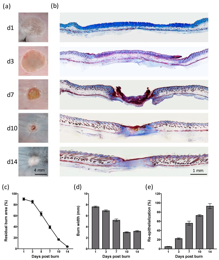
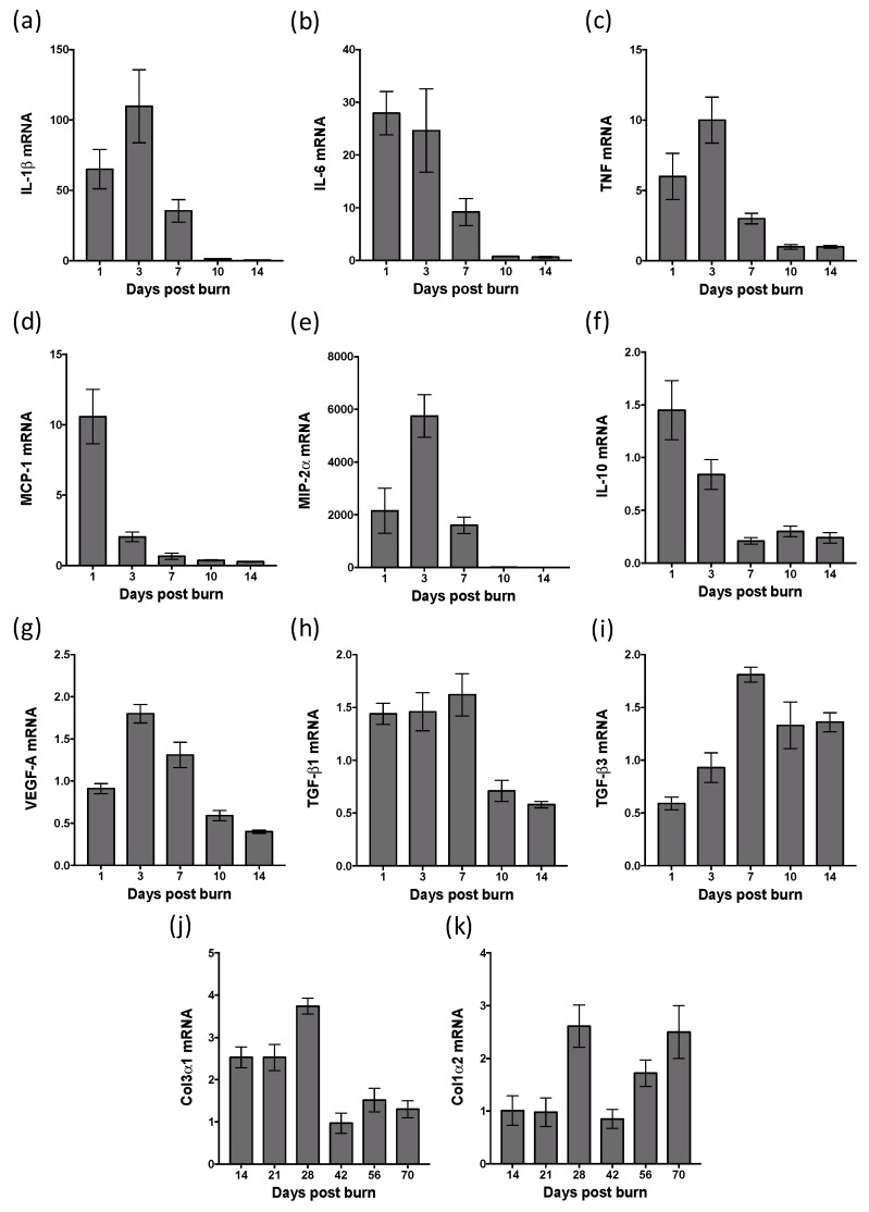
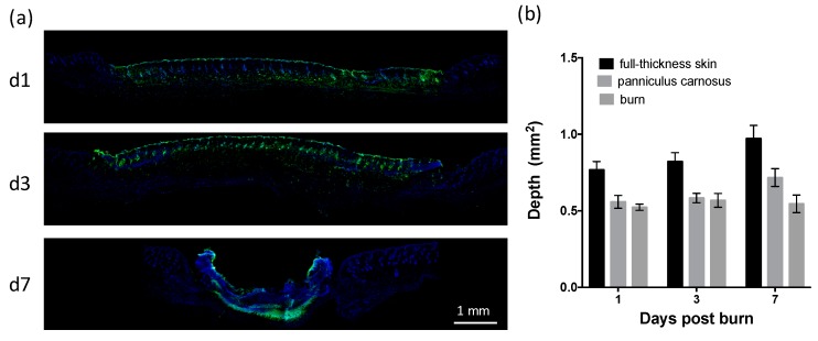
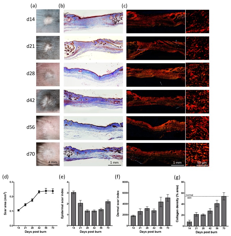
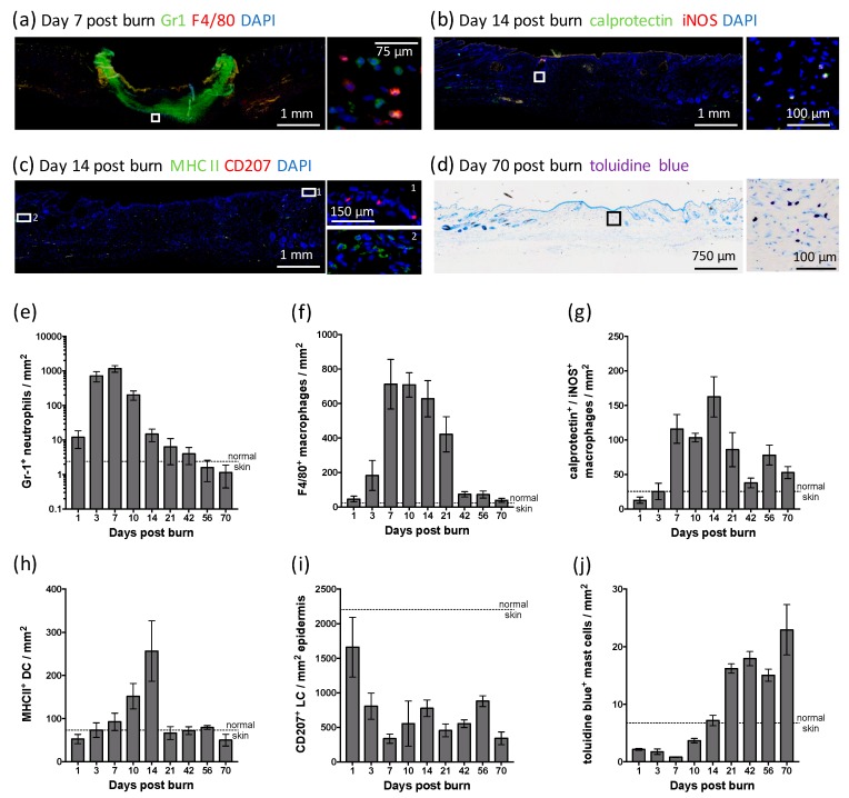
Many burn interventions aim to target the inflammatory response as a means of enhancing healing or limiting hypertrophic scarring. Murine models of human burns have been developed, but the inflammatory response to injury in these models has not been well defined. The aim of this study was to profile inflammatory cell populations and gene expression relative to healing and scarring in a murine model of thermal burns. Cutaneous injuries were created on the dorsal region of C57Bl/6 mice using a heated metal rod. Animals were euthanized at selected time points over ten weeks, with the lesions evaluated using macroscopic measurements, histology, immunofluorescent histochemistry and quantitative PCR. The burn method generated a reproducible, partial-thickness injury that healed within two weeks through both contraction and re-epithelialization, in a manner similar to human burns. The injury caused an immediate increase in pro-inflammatory cytokine and chemokine expression, coinciding with an influx of neutrophils, and the disappearance of Langerhans cells and mast cells. This preceded an influx of dendritic cells and macrophages, a quarter of which displayed an inflammatory (M1) phenotype, with both populations peaking at closure. As with human burns, the residual scar increased in size, epidermal and dermal thickness, and mast cell numbers over 10 weeks, but abnormal collagen I-collagen III ratios, fibre organization and macrophage populations resolved 3⁻4 weeks after closure. Characterisation of the inflammatory response in this promising murine burn model will assist future studies of burn complications and aid in the preclinical testing of new anti-inflammatory and anti-scarring therapies.
许多烧伤干预措施旨在靶向炎症反应,以促进愈合或限制增生性瘢痕形成。已经开发出了人类烧伤的小鼠模型,但这些模型中损伤的炎症反应尚未得到很好的定义。本研究的目的是在小鼠热烧伤模型中,对与愈合和瘢痕形成相关的炎症细胞群和基因表达进行分析。使用加热的金属棒在 C57Bl/6 小鼠的背部区域创建皮肤损伤。在十周的时间内选择时间点处死动物,并使用宏观测量、组织学、免疫荧光组织化学和定量 PCR 评估病变。烧伤方法产生了一种可重复的、部分厚度的损伤,通过收缩和再上皮化在两周内愈合,这与人类烧伤相似。损伤会立即引起促炎细胞因子和趋化因子表达增加,伴随着中性粒细胞的涌入,朗格汉斯细胞和肥大细胞的消失。这发生在树突状细胞和巨噬细胞的涌入之前,其中四分之一表现出炎症(M1)表型,这两种细胞群在闭合时达到峰值。与人类烧伤一样,残余瘢痕在 10 周内会增加大小、表皮和真皮厚度以及肥大细胞数量,但异常的胶原 I-胶原 III 比值、纤维组织和巨噬细胞群在闭合后 3-4 周得到解决。对这种有前景的小鼠烧伤模型中炎症反应的特征描述将有助于未来对烧伤并发症的研究,并有助于新的抗炎和抗瘢痕形成治疗的临床前测试。