Department of Applied Physics and Science for Life Laboratory, KTH Royal Institute of Technology, 100 44, Stockholm, Sweden.
Molecular Microscopy and Spectroscopy, Istituto Italiano di Tecnologia, Via Morego 30, 16136, Genoa, Italy.
Nat Commun. 2019 Feb 1;10(1):556. doi: 10.1038/s41467-019-08442-4.
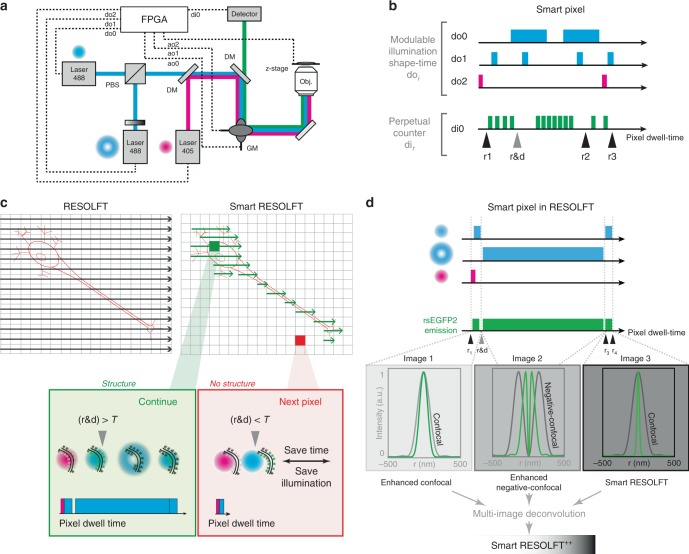
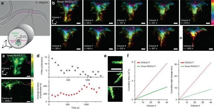
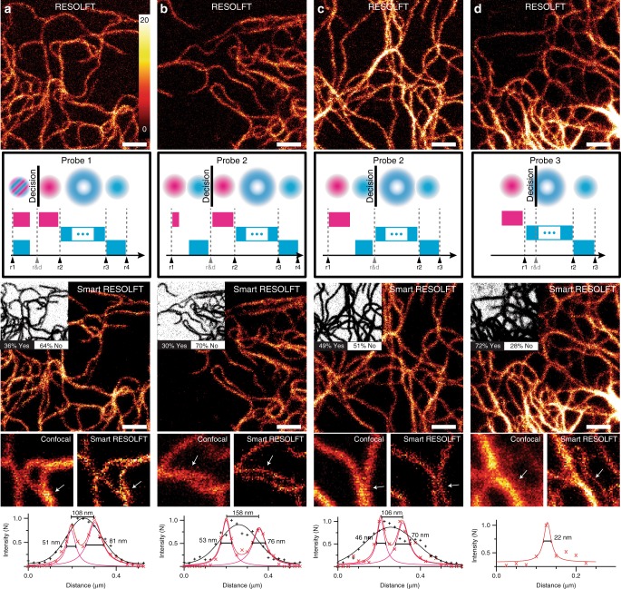
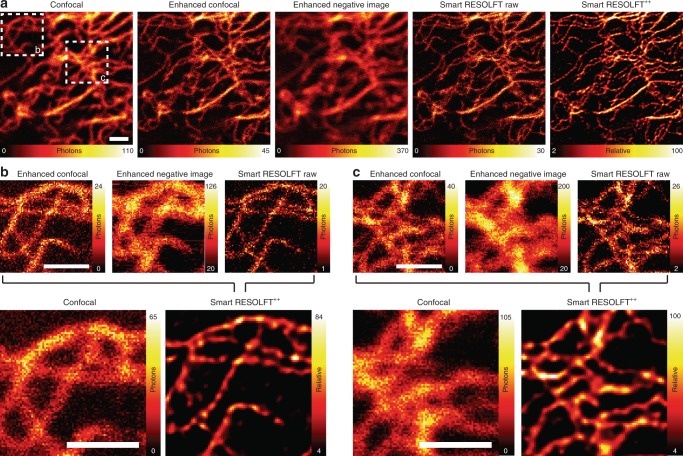
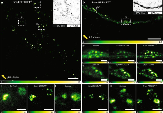
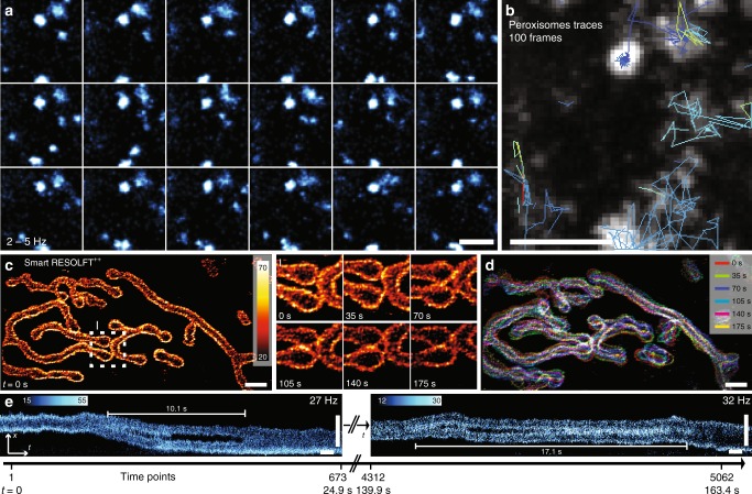
RESOLFT fluorescence nanoscopy can nowadays image details far beyond the diffraction limit. However, signal to noise ratio (SNR) and temporal resolution are still a concern, especially deep inside living cells and organisms. In this work, we developed a non-deterministic scanning approach based on a real-time feedback system which speeds up the acquisition up to 6-fold and decreases the light dose by 70-90% for in vivo imaging. Also, we extended the information content of the images by acquiring the complete temporal evolution of the fluorescence generated by reversible switchable fluorescent proteins. This generates a series of images with different spatial resolution and SNR, from conventional to RESOLFT images, which combined through a multi-image deconvolution algorithm further enhances the effective resolution. We reported nanoscale imaging of organelles up to 35 Hz and actin dynamics during an invasion process at a depth of 20-30 µm inside a living Caenorhabditis elegans worm.
RESOLFT 荧光纳米显微镜如今可以对远超出衍射极限的细节进行成像。然而,信噪比(SNR)和时间分辨率仍然是一个关注点,尤其是在活细胞和生物体内深处。在这项工作中,我们开发了一种基于实时反馈系统的非确定性扫描方法,该方法将采集速度提高了 6 倍,并将活体内成像的光剂量降低了 70-90%。此外,我们通过获取可逆可切换荧光蛋白产生的荧光的完整时间演化,扩展了图像的信息量。这生成了一系列具有不同空间分辨率和 SNR 的图像,从传统 RESOLFT 图像到 RESOLFT 图像,通过多图像反卷积算法对其进行组合,进一步提高了有效分辨率。我们报告了在活体秀丽隐杆线虫体内 20-30µm 深处,对细胞器进行的高达 35Hz 的纳米尺度成像,以及在入侵过程中肌动蛋白动力学的成像。