Department of Applied Physics and Science for Life Laboratory, KTH Royal Institute of Technology, Stockholm, Sweden.
Nat Methods. 2022 Oct;19(10):1268-1275. doi: 10.1038/s41592-022-01588-y. Epub 2022 Sep 8.
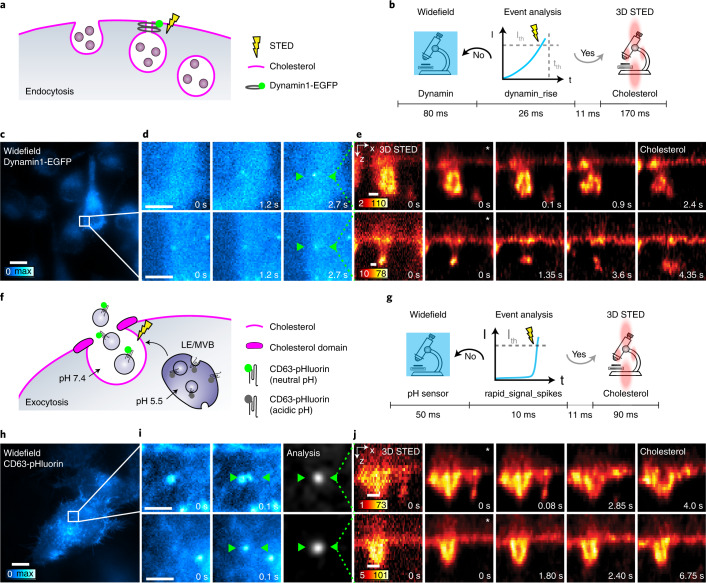
Monitoring the proteins and lipids that mediate all cellular processes requires imaging methods with increased spatial and temporal resolution. STED (stimulated emission depletion) nanoscopy enables fast imaging of nanoscale structures in living cells but is limited by photobleaching. Here, we present event-triggered STED, an automated multiscale method capable of rapidly initiating two-dimensional (2D) and 3D STED imaging after detecting cellular events such as protein recruitment, vesicle trafficking and second messengers activity using biosensors. STED is applied in the vicinity of detected events to maximize the temporal resolution. We imaged synaptic vesicle dynamics at up to 24 Hz, 40 ms after local calcium activity; endocytosis and exocytosis events at up to 11 Hz, 40 ms after local protein recruitment or pH changes; and the interaction between endosomal vesicles at up to 3 Hz, 70 ms after approaching one another. Event-triggered STED extends the capabilities of live nanoscale imaging, enabling novel biological observations in real time.
监测介导所有细胞过程的蛋白质和脂质需要具有更高空间和时间分辨率的成像方法。STED(受激发射损耗)纳米显微镜能够快速成像活细胞中的纳米级结构,但受到光漂白的限制。在这里,我们提出了事件触发的 STED,这是一种自动化的多尺度方法,能够在使用生物传感器检测到细胞事件(如蛋白质募集、囊泡运输和第二信使活性)后,快速启动二维(2D)和 3D STED 成像。STED 应用于检测到的事件附近,以最大限度地提高时间分辨率。我们以高达 24 Hz、局部钙活动后 40 毫秒的速度成像突触囊泡动力学;以高达 11 Hz、局部蛋白质募集或 pH 变化后 40 毫秒的速度成像内吞作用和胞吐作用事件;以及以高达 3 Hz、彼此接近后 70 毫秒的速度成像内体囊泡之间的相互作用。事件触发的 STED 扩展了活纳米尺度成像的功能,能够实时进行新的生物学观察。