Koyanagi Yuki, Kubo Chiaki, Nagata Shigenori, Ryu Ayumi, Hatano Koji, Kano Rieko, Tanada Satoshi, Ashimura Jun-Ichi, Idota Atsushi, Kamiura Shoji, Yamasaki Tomoyuki, Nakatsuka Shin-Ichi
Department of Clinical Laboratory, Osaka International Cancer Institute Hospital, Osaka, Japan.
Department of Diagnostic Pathology and Cytology, Osaka International Cancer Institute Hospital, 3-1-69 Otemae, Chuo-ku, Osaka, 541-8567, Japan.
Diagn Pathol. 2019 Feb 2;14(1):9. doi: 10.1186/s13000-019-0788-2.
Pagetoid spread of urothelial carcinoma (UC) to the lower genital tract is quite a rare and diagnostically challenging condition. Pagetoid urothelial intraepithelial neoplasia extending to the vagina is difficult to diagnose, especially in remote recurrences without symptomatic or macroscopic lesions typical to Paget disease. However, its identification by cervical screening cytology is important because UC is often characterized by a long history of relapse.
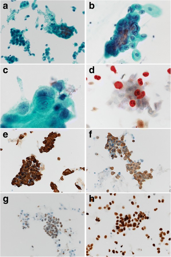
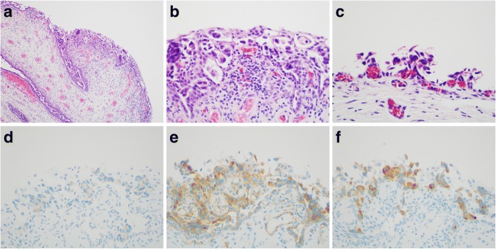
A 68-year-old Japanese postmenopausal woman developed brown vaginal discharge after radical cystectomy for bladder cancer (high-grade UC, pT2a pN0 cM0 [Union for International Cancer Control, 8th edition]) concomitant with focal in-situ UC in the urethra. She had a history of left renal pelvis UC, which was surgically removed 9 months before the radical cystectomy. Gynecologic examination of the lower genital tract was unremarkable although cervical screening cytology demonstrated severely atypical cells with pleomorphism repeatedly. Cervical colposcopy and diagnostic conization revealed no cervical neoplasm. In retrospect, immunocytochemical p16/Ki-67 dual staining for the previous cervical screening was negative for p16 labeling, and the neoplastic cells were positive for cytokeratins 7 and 20, p63, and GATA binding protein 3. No high-risk human papillomavirus genotype was identified by an automated DNA chip system using liquid-based cytology samples. Eleven months post-cystectomy, punch biopsy of the vulva and vagina confirmed intraepithelial UC in the juxtaposed squamous epithelium with pagetoid spread demonstrating positivity for specific urothelial markers: uroplakins II and III and thrombomodulin. Concurrent invasive malignancy was ruled out, and CO laser vaporization of the vulvar and vaginal lesion was performed. The patient remained alive without evidence of invasive malignancy for 14 months after the radical cystectomy for bladder cancer.
To detect recurrent pagetoid urothelial intraepithelial neoplasia with pagetoid spread in the lower genital tract, pathologists should recognize the history of prior UC with special attention to absence of p16 labeling in cervical cytology as a pointer to the diagnosis of urothelial cancer. Using further biopsy and immunohistochemical confirmation of UC relapse, investigation to rule out invasive malignancies and careful follow-up throughout the patient's lifetime is recommended.
尿路上皮癌(UC)向低生殖道的派杰样扩散是一种非常罕见且诊断具有挑战性的情况。派杰样尿路上皮内瘤变累及阴道很难诊断,尤其是在没有派杰病典型症状或肉眼可见病变的远处复发时。然而,通过宫颈筛查细胞学识别它很重要,因为UC通常具有复发史较长的特点。
一名68岁的日本绝经后女性在因膀胱癌(高级别UC,pT2a pN0 cM0 [国际癌症控制联盟第8版])行根治性膀胱切除术时,伴有尿道局灶原位UC,术后出现褐色阴道分泌物。她有左肾盂UC病史,在根治性膀胱切除术9个月前已手术切除。尽管宫颈筛查细胞学多次显示有严重异形的非典型细胞,但对低生殖道的妇科检查无异常。宫颈阴道镜检查和诊断性锥切未发现宫颈肿瘤。回顾既往宫颈筛查的免疫细胞化学p16/Ki-67双重染色,p16标记为阴性,肿瘤细胞细胞角蛋白7和20、p63及GATA结合蛋白3呈阳性。使用液基细胞学样本的自动DNA芯片系统未检测到高危人乳头瘤病毒基因型。膀胱切除术后11个月,对外阴和阴道进行活检,证实并列鳞状上皮内有上皮内UC伴派杰样扩散,显示特定尿路上皮标志物尿膜素II和III及血栓调节蛋白呈阳性。排除了同时存在的浸润性恶性肿瘤,并对外阴和阴道病变进行了CO2激光汽化术。在因膀胱癌行根治性膀胱切除术后14个月,患者存活,无浸润性恶性肿瘤证据。
为了检测低生殖道中伴有派杰样扩散的复发性派杰样尿路上皮内瘤变,病理学家应了解既往UC病史,特别注意宫颈细胞学中p16标记缺失这一提示尿路上皮癌诊断的指标。建议通过进一步活检和免疫组化确认UC复发,排除浸润性恶性肿瘤,并在患者的整个生命周期内进行仔细随访。