Huang Zhiang, Huang Haidong, Ning Yunye, Han Jin, Shen Yibo, Shi Hui, Wang Qin, Bai Chong, Li Qiang, Michael Simoff, Zarogoulidis Paul, Hohenforst-Schmidt Wolfgang, Konstantinou Fotis, Turner J Francis, Koulouris Charilaos, Katsaounis Athanasios, Amaniti Aikaterini, Mantalovas Stylianos, Pavlidis Efstathios, Giannakidis Dimitrios, Passos Ioannis, Michalopoulos Nikolaos, Kosmidis Christoforos, Mogoantă Stelian Ştefăniţă, Sapalidis Konstantinos
Department of Respiratory & Critical Care Medicine, Changhai Hospital, the Second Military Medical University, Shanghai, 200433, China.
Department of Respiratory, The First Affiliated Hospital of Henan University, Henan Kaifeng, 475000, China.
J Cancer. 2019 Jan 1;10(3):634-642. doi: 10.7150/jca.28755. eCollection 2019.
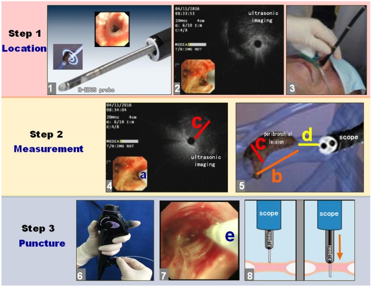
The diagnosis of peribronchial pulmonary lesions located in the tertiary bronchi, also known as segmental bronchi, as well as, the 4th order and 5th order segmental bronchi is very difficult. Histopathological specimens cannot be easily obtained by endobronchial biopsies (EBBX) due to the patent but small segmental bronchial lumen. The aim of the present study was to evaluate the diagnostic accuracy and safety of the novel technique with radial probe endobronchial ultrasound (R-EBUS) assisted conventional transbronchial needle aspiration (C-TBNA) in the diagnosis of solitary peribronchial pulmonary lesions located in segmental bronchi from 3th to 5th order. From December 2014 to December 2015, 16 patients with solitary peribronchial pulmonary lesions in the segmental bronchi from 3th to 5th order confirmed by computed tomography (CT) were enrolled. The lesions were located using radial probe endobronchial ultrasound (R-EBUS) to determine the sites of conventional transbronchial needle aspiration (C-TBNA), then, histopathological specimens were obtained using the technique of C-TBNA. The final pathological diagnosis was made based on the findings from the surgical specimens. Statistical analyses were performed for specimen results and complications. On pathological evaluation, 14 of the 16 specimens were malignant, including 8 adenocarcinomas, 4 squamous cell carcinomas, and 2 small cell carcinomas, while 2 were non-malignant diseases. The diagnostic accuracy rate, sensitivity and missed diagnosis rates were 87.5%, 87.5% and 12.5%, respectively. When Combined the results of cytology with histologic samples obtained from C-TBNA the total diagnostic accuracy rate, sensitivity and missed diagnosis rate were 93.75%, 93.75% and 6.25%, respectively. There were 2 cases of bleeding complications >5 mL after C-TBNA, and both were resolved with endobronchial management. The combination of R-EBUS with C-TBNA was advantageous and safe for the diagnosis of solitary peribronchial pulmonary lesions located in the segmental bronchi. However, possible bleeding complications should be anticipated with needle aspiration. Further verification of this combined application should be investigated in larger clinical trials.
诊断位于三级支气管(也称为肺段支气管)以及第四级和第五级肺段支气管的支气管周围肺部病变非常困难。由于肺段支气管管腔虽通畅但狭小,通过支气管内活检(EBBX)不易获取组织病理学标本。本研究的目的是评估采用径向探头支气管内超声(R-EBUS)辅助传统经支气管针吸活检(C-TBNA)新技术对位于第三级至第五级肺段支气管的孤立性支气管周围肺部病变的诊断准确性和安全性。2014年12月至2015年12月,纳入16例经计算机断层扫描(CT)确诊的位于第三级至第五级肺段支气管的孤立性支气管周围肺部病变患者。使用径向探头支气管内超声(R-EBUS)定位病变,以确定传统经支气管针吸活检(C-TBNA)的部位,然后采用C-TBNA技术获取组织病理学标本。最终病理诊断基于手术标本的检查结果。对标本结果和并发症进行统计分析。病理评估显示,16例标本中有14例为恶性,包括8例腺癌、4例鳞状细胞癌和2例小细胞癌,2例为非恶性疾病。诊断准确率、灵敏度和漏诊率分别为87.5%、87.5%和12.5%。将C-TBNA的细胞学结果与组织学样本结果相结合时,总诊断准确率、灵敏度和漏诊率分别为93.75%、93.75%和6.25%。C-TBNA术后有2例出血并发症出血量>5 mL,均通过支气管内处理得以解决。R-EBUS与C-TBNA联合应用对诊断位于肺段支气管的孤立性支气管周围肺部病变是有效且安全的。然而,针吸活检时应预见到可能的出血并发症。这种联合应用的进一步验证应在更大规模的临床试验中进行研究。