China-USA Neuroimaging Research Institute, Department of Radiology of the Second Affiliated Hospital and Yuying Children's Hospital, Wenzhou Medical University, Wenzhou, Zhejiang 325027, China.
The Eye Hospital, Wenzhou Medical University, Wenzhou, Zhejiang 325027, China.
Neural Plast. 2019 Jan 8;2019:2981764. doi: 10.1155/2019/2981764. eCollection 2019.
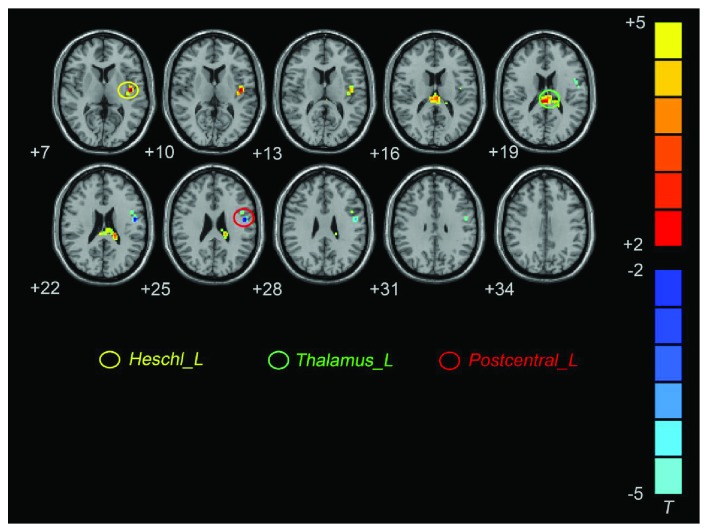
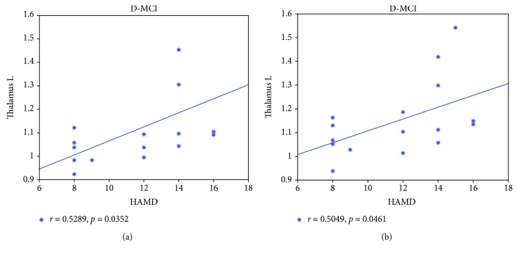
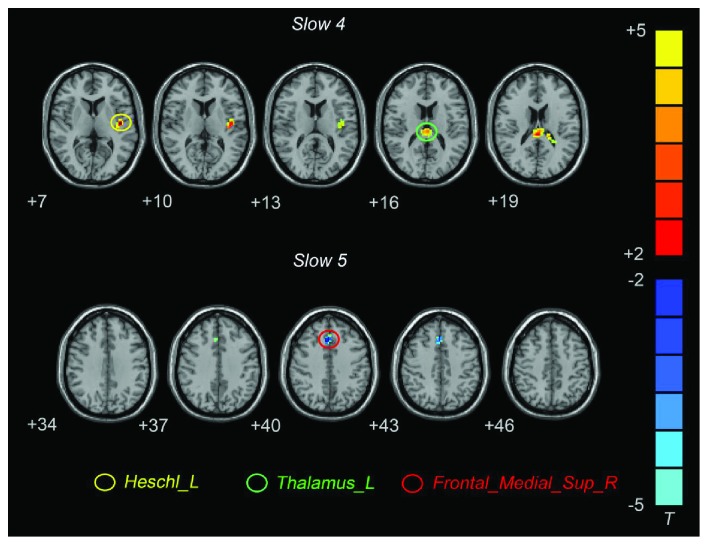
Depressive symptoms are common in individuals with mild cognitive impairment (MCI) who have an increased risk of dementia. It is currently unclear whether the pattern of spontaneous brain activity in patients with MCI differs between subjects with and without depressive symptoms. The current study sought to investigate the features of spontaneous brain activity in MCI patients with depressive symptoms (D-MCI) using coherence regional homogeneity (CReHo) analysis with resting-state functional magnetic resonance imaging (rsfMRI). We obtained rsfMRI data in 16 MCI patients with depressive symptoms and 18 nondepressed MCI patients (nD-MCI) using a 3 T scanner. Statistical analyses were performed to determine the regions in which ReHo differed between the two groups in specific frequency bands, slow-4 (0.027-0.073 Hz) and slow-5 (0.010-0.027 Hz), and typical bands (0.01-0.08 Hz). Correlation analyses were performed between the CReHo index of these regions and clinical variables to evaluate the relationship between CReHo and pathophysiological measures in the two groups. Our results showed that D-MCI patients exhibited significantly higher CReHo in the left Heschl's gyrus and left thalamus and lower CReHo in the left postcentral gyrus in the typical frequency band. In the slow-4 frequency band, D-MCI patients showed significantly higher CReHo in the left Heschl's gyrus and left thalamus. In the slow-5 frequency band, D-MCI patients exhibited significantly lower CReHo in the superior medial prefrontal gyrus. In addition, the results revealed that CReHo values in the left thalamus were positively correlated with Hamilton Depression Rating Scale (HAMD) scores in D-MCI patients. These results suggest that the sensorimotor network may be one of the main pathophysiological factors in D-MCI.
抑郁症状在轻度认知障碍 (MCI) 患者中很常见,而这些患者痴呆的风险增加。目前尚不清楚 MCI 患者的自发性大脑活动模式是否在有无抑郁症状的患者之间存在差异。本研究旨在使用静息态功能磁共振成像 (rsfMRI) 中的相干区域同质性 (CReHo) 分析来研究有抑郁症状的 MCI 患者 (D-MCI) 的自发性大脑活动特征。我们使用 3T 扫描仪获得了 16 名有抑郁症状的 MCI 患者和 18 名无抑郁 MCI 患者 (nD-MCI) 的 rsfMRI 数据。统计分析用于确定两组在特定频带(慢-4(0.027-0.073Hz)和慢-5(0.010-0.027Hz)和典型频带(0.01-0.08Hz)中 ReHo 不同的区域。对这些区域的 CReHo 指数与临床变量进行相关性分析,以评估两组中 CReHo 与病理生理测量值之间的关系。我们的结果表明,D-MCI 患者在典型频率带中左侧 Heschl 回和左侧丘脑的 CReHo 明显升高,左侧中央后回的 CReHo 明显降低。在慢-4 频带中,D-MCI 患者左侧 Heschl 回和左侧丘脑的 CReHo 明显升高。在慢-5 频带中,D-MCI 患者的上内侧前额叶的 CReHo 明显降低。此外,结果表明 D-MCI 患者左侧丘脑的 CReHo 值与汉密尔顿抑郁量表 (HAMD) 评分呈正相关。这些结果表明感觉运动网络可能是 D-MCI 的主要病理生理因素之一。