Schlegel Research Institute for Aging, Department of Electrical & Computer Engineering, University of Waterloo, Waterloo, ON, N2L 3G1, Canada.
Bioprober Corporation, Seattle, WA, 98004, USA.
Med Phys. 2019 Apr;46(4):1620-1633. doi: 10.1002/mp.13437. Epub 2019 Mar 4.
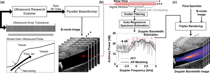
Flow instability has been shown to contribute to the risk of future cardiovascular and cerebrovascular events. Nonetheless, it is challenging to noninvasively detect and identify flow instability in blood vessels. Here, we present a new framework called Doppler ultrasound bandwidth imaging (DUBI) that uses high-frame-rate ultrasound and Doppler bandwidth analysis principles to assess flow instability within an image view.
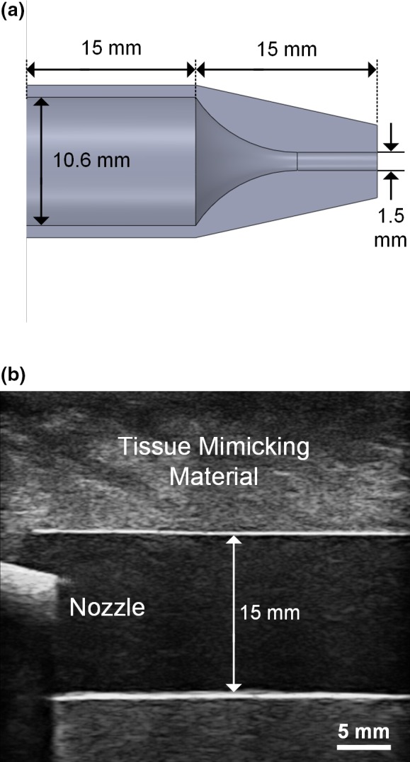
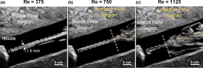
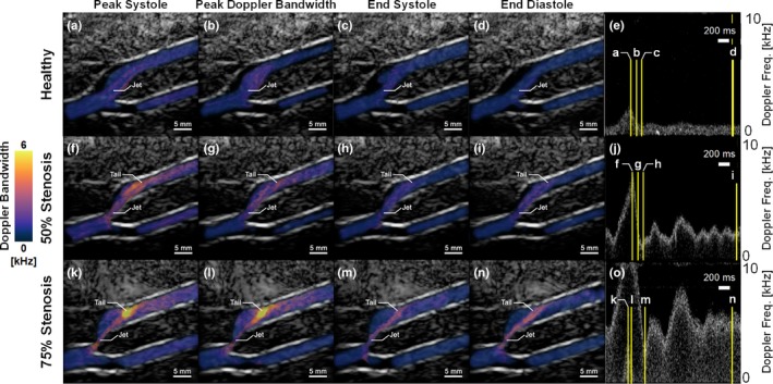
Doppler ultrasound bandwidth imaging seeks to estimate the instantaneous Doppler bandwidth based on autoregressive modeling at every pixel position of data frames acquired from high-frame-rate plane wave pulsing. This new framework is founded upon the principle that flow instability naturally gives rise to a wide range of flow velocities over a sample volume, and such velocity range in turn yields a larger Doppler bandwidth estimate. The ability for DUBI to map unstable flow was first tested over a range of fluid flow conditions (ranging from laminar to turbulent) with a nozzle-flow phantom. As a further demonstration, DUBI was applied to assess flow instability in healthy and stenosed carotid bifurcation phantoms.
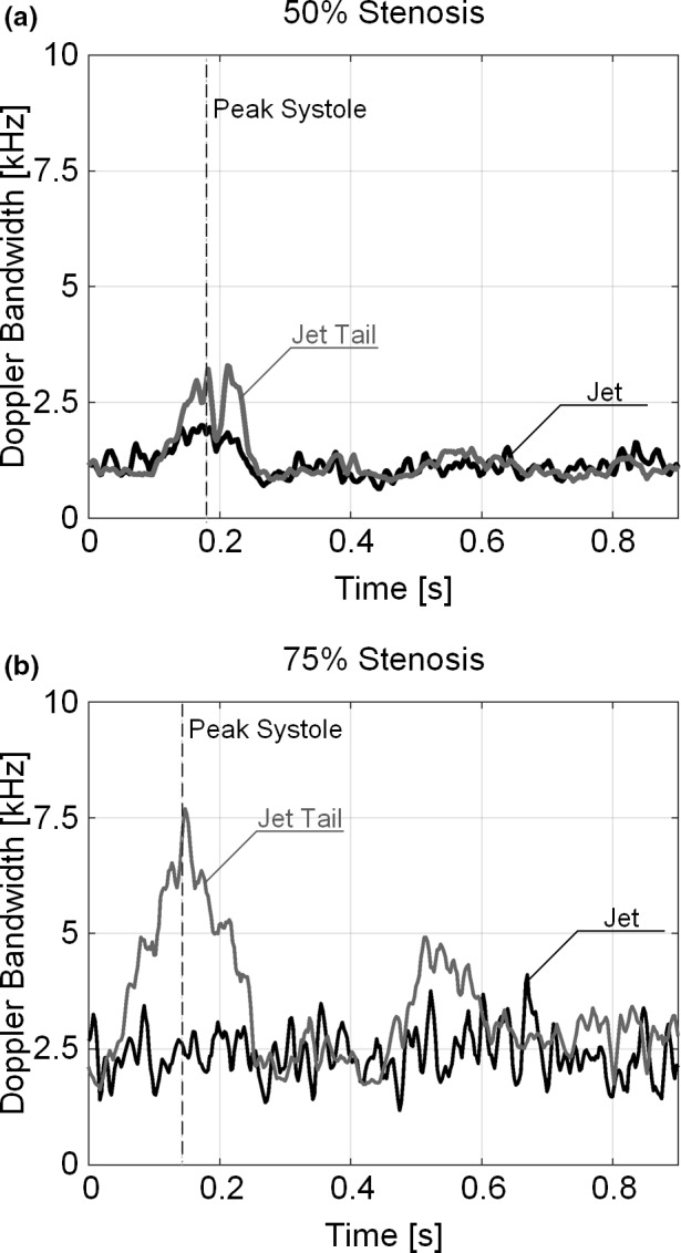
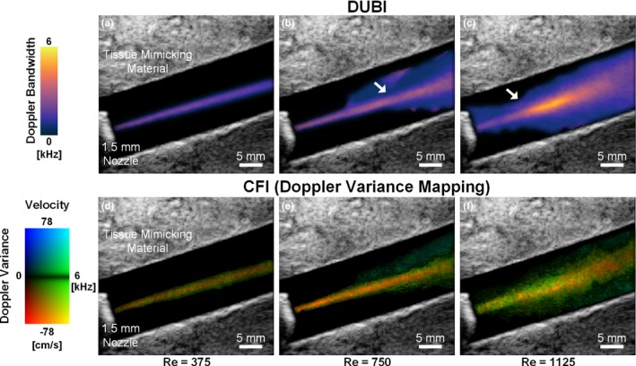
Nozzle-flow phantom results showed that DUBI can effectively detect and visualize the difference in Doppler bandwidth magnitude (increased from 2.1 to 5.2 kHz) at stable and unstable flow regions in an image view. Receiver operating characteristic analysis also showed that DUBI can achieve optimal sensitivity and specificity of 0.72 and 0.83, respectively. In the carotid phantom experiments, differences were observed in the spatiotemporal dynamics of Doppler bandwidth over a cardiac cycle. Specifically, as the degree of stenosis increased (from 50% to 75%), DUBI showed an increase in Doppler bandwidth magnitude from 1.4 kHz in the healthy bifurcation to 7.7 kHz at the jet tail located downstream from a 75% stenosis site, thereby indicating flow perturbation in the stenosed bifurcations.
DUBI can detect unstable flow. This new technique can provide useful hemodynamic information that may aid clinical diagnosis of atherosclerosis.
血流不稳定性已被证明会增加未来心血管和脑血管事件的风险。尽管如此,非侵入性地检测和识别血管内的血流不稳定性仍然具有挑战性。在这里,我们提出了一种新的框架,称为多普勒超声带宽成像(DUBI),它使用高帧率超声和多普勒带宽分析原理来评估图像视图内的血流不稳定性。
多普勒超声带宽成像试图根据高帧率平面波脉冲采集的数据帧中每个像素位置的自回归建模来估计瞬时多普勒带宽。该新框架基于这样的原理:血流不稳定性自然会导致样本体积内的流速范围很宽,而这种速度范围反过来又会产生更大的多普勒带宽估计值。首先,通过喷嘴流幻影在一系列流体流动条件(从层流到湍流)下测试 DUBI 对不稳定流动的映射能力。作为进一步的演示,将 DUBI 应用于评估健康和狭窄颈动脉分叉幻影中的血流不稳定性。
喷嘴流幻影结果表明,DUBI 可以有效地检测和可视化图像视图中稳定和不稳定流动区域的多普勒带宽幅度(从 2.1 增加到 5.2 kHz)的差异。接收器操作特性分析还表明,DUBI 可以分别达到 0.72 和 0.83 的最佳灵敏度和特异性。在颈动脉幻影实验中,在心动周期内观察到多普勒带宽的时空动力学差异。具体来说,随着狭窄程度的增加(从 50%增加到 75%),DUBI 显示出从健康分叉处的 1.4 kHz 到位于 75%狭窄部位下游的射流尾部处的 7.7 kHz 的多普勒带宽幅度增加,从而表明狭窄分叉处的血流扰动。
DUBI 可以检测到不稳定的流动。这项新技术可以提供有用的血液动力学信息,可能有助于动脉粥样硬化的临床诊断。