Neuroscience Program and Department of Physiology, Michigan State University, 567 Wilson Road, BPS 3182, East Lansing, MI, 48824, USA.
Mol Brain. 2019 Feb 8;12(1):10. doi: 10.1186/s13041-019-0435-6.
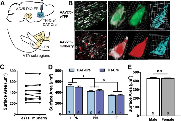
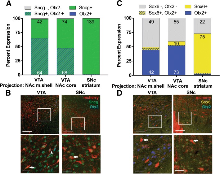
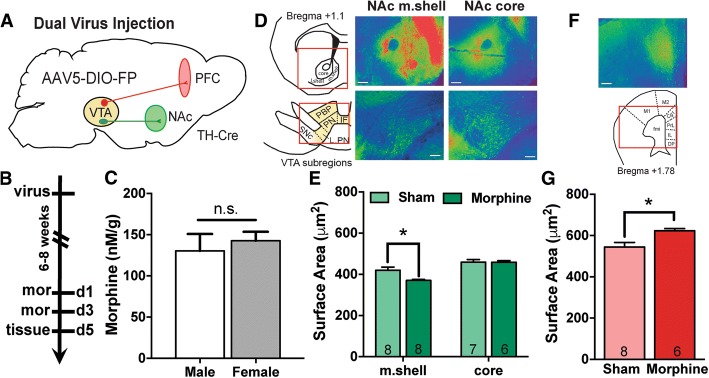
Chronic opiate exposure induces neuroadaptations in the mesocorticolimbic system including ventral tegmental area (VTA) dopamine (DA) neurons, whose soma size is decreased following opiate exposure. Yet it is now well documented that VTA DA neurons are heterogeneous, with notable differences between VTA DA neurons based on their projection target. Therefore, we sought to determine whether chronic morphine induced similar changes in the morphology of VTA DA neurons that project to the nucleus accumbens (NAc) and prefrontal cortex (PFC). We utilized Cre-dependent retrograde viral vectors in DA Cre driver lines to label VTA DA neurons that projected to NAc and PFC and assessed neuronal soma size. Consistent with previous data, the soma size of VTA DA neurons that projected to the NAc medial shell was decreased following morphine exposure. However, soma size of VTA DA neurons that projected to the NAc core was unaltered by morphine. Interestingly, morphology of PFC-projecting VTA DA neurons was also altered by morphine, but in this case soma size was increased compared to sham controls. Differences in basal soma size were also noted, suggesting stable differences in projection-specific morphology in addition to drug-induced changes. Together, these data suggest morphine-induced changes in VTA DA morphology occur within distinct VTA DA populations and that study of opiate-induced structural plasticity of individual VTA DA subcircuits may be critical for understanding addiction-related behavior.
慢性阿片类药物暴露会导致中脑边缘多巴胺(DA)神经元发生神经适应性改变,包括腹侧被盖区(VTA)DA 神经元,其细胞体大小在阿片类药物暴露后会减小。然而,现在有充分的证据表明,VTA DA 神经元是异质的,根据其投射目标,VTA DA 神经元之间存在显著差异。因此,我们试图确定慢性吗啡是否会对投射到伏隔核(NAc)和前额叶皮层(PFC)的 VTA DA 神经元的形态产生类似的变化。我们利用 Cre 依赖性逆行病毒载体在 DA Cre 驱动系中标记投射到 NAc 和 PFC 的 VTA DA 神经元,并评估神经元细胞体大小。与先前的数据一致,投射到 NAc 内侧壳的 VTA DA 神经元的细胞体大小在吗啡暴露后减小。然而,投射到 NAc 核心的 VTA DA 神经元的细胞体大小不受吗啡影响。有趣的是,吗啡也改变了投射到 PFC 的 VTA DA 神经元的形态,但在这种情况下,与假手术对照组相比,细胞体大小增加。还注意到基础细胞体大小的差异,这表明除了药物诱导的变化外,投射特异性形态还存在稳定的差异。总之,这些数据表明,吗啡诱导的 VTA DA 形态变化发生在不同的 VTA DA 神经元群体中,研究单个 VTA DA 亚电路的阿片类药物诱导的结构可塑性对于理解与成瘾相关的行为可能至关重要。