• 文献检索
  • 文档翻译
  • 深度研究
  • 学术资讯
  • Suppr Zotero 插件Zotero 插件
  • 邀请有礼
  • 套餐&价格
  • 历史记录
应用&插件
Suppr Zotero 插件Zotero 插件浏览器插件Mac 客户端Windows 客户端微信小程序
定价
高级版会员购买积分包购买API积分包
服务
文献检索文档翻译深度研究API 文档MCP 服务
关于我们
关于 Suppr公司介绍联系我们用户协议隐私条款
关注我们

Suppr 超能文献

核心技术专利:CN118964589B侵权必究
粤ICP备2023148730 号-1Suppr @ 2026

文献检索

告别复杂PubMed语法,用中文像聊天一样搜索,搜遍4000万医学文献。AI智能推荐,让科研检索更轻松。

立即免费搜索

文件翻译

保留排版,准确专业,支持PDF/Word/PPT等文件格式,支持 12+语言互译。

免费翻译文档

深度研究

AI帮你快速写综述,25分钟生成高质量综述,智能提取关键信息,辅助科研写作。

立即免费体验

大肠杆菌外膜囊泡诱导肠上皮Caco-2细胞中的DNA双链断裂。

Escherichia Coli Outer Membrane Vesicles Induced DNA Double-Strand Breaks in Intestinal Epithelial Caco-2 Cells.

作者信息

Ling Zhou, Dayong Chen, Denggao Yu, Yiting Wang, Liaoqiong Fang, Zhibiao Wang

机构信息

State Key Laboratory of Ultrasound Engineering in Medicine Co-Founded by Chongqing and the Ministry of Science and Technology, College of Biomedical Engineering, Chongqing Medical University, Chongqing, China (mainland).

出版信息

Med Sci Monit Basic Res. 2019 Feb 11;25:45-52. doi: 10.12659/MSMBR.913756.

DOI:10.12659/MSMBR.913756
PMID:30739907
原文链接:https://pmc.ncbi.nlm.nih.gov/articles/PMC6381811/
Abstract

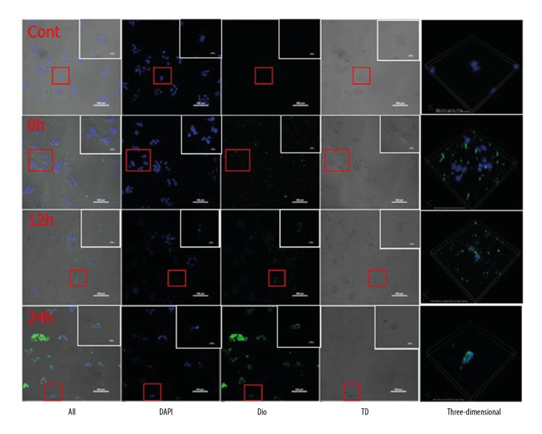
BACKGROUND Recent studies have shown that Escherichia coli induced digestive tract diseases may be related to outer membrane vesicles (OMVs) induced intestinal double-strand breaks (DSBs) in intestinal epithelial cells. This study aimed to compare the impact of OMVs forces on DSBs in intestinal epithelial Caco-2 cells, and provide a new treatment for digestive diseases caused by E. coli. MATERIAL AND METHODS E.coli OMVs were prepared and co-cultured with Caco-2 cells. The uptake of OMVs by Caco-2 cells was observed by confocal microscopy. The γ-H2AX protein was detected by western-blots. The DSBs caused by OMVs was detected by single cell gel electrophoresis. RESULTS The particle size analyzer showed that the average diameters of OMVs centrifuged at 20 000×g and 50 000×g were 217.5±7.29 nm and 186.3±6.59 nm (P<0.05), respectively. Transmission electron microscopy of the OMVs revealed a lipid bilayer structure with a variety of different sizes. Confocal fluorescence microscopy revealed that OMVs almost completely entered Caco-2 cells after 24 hours. The ratio of γ-H2AX protein band gray value normalized data in the OMVs centrifuged at 20 000×g and 50 000×g, and the control group (without OMVs) were 2.23±0.18, 1.58±0.20, 1±0.30 (P<0.05), respectively, while DNA levels of the comet tail (TailDNA%, TDNA%) were 72.21±14.61%, 23.11±4.98%, and 1.02±1.41% (P<0.05), respectively. The corresponding DNA damage was categorized as high (grade 3), moderate (grade 2), and no damage (grade 0). CONCLUSIONS Different sizes of OMVs induced different degrees of DNA damage in intestinal epithelial Caco-2 cells.

摘要

背景 近期研究表明,大肠杆菌引起的消化道疾病可能与外膜囊泡(OMVs)诱导肠上皮细胞中的肠道双链断裂(DSBs)有关。本研究旨在比较OMVs对肠上皮Caco-2细胞中DSBs的影响,并为大肠杆菌引起的消化系统疾病提供新的治疗方法。 材料与方法 制备大肠杆菌OMVs并与Caco-2细胞共培养。通过共聚焦显微镜观察Caco-2细胞对OMVs的摄取。通过蛋白质免疫印迹法检测γ-H2AX蛋白。通过单细胞凝胶电泳检测由OMVs引起的DSBs。 结果 粒度分析仪显示,在20000×g和50000×g离心的OMVs的平均直径分别为217.5±7.29nm和186.3±6.59nm(P<0.05)。OMVs的透射电子显微镜显示具有各种不同大小的脂质双层结构。共聚焦荧光显微镜显示,24小时后OMVs几乎完全进入Caco-2细胞。在20000×g和50000×g离心的OMVs以及对照组(无OMVs)中,γ-H2AX蛋白条带灰度值归一化数据的比例分别为2.23±0.18、1.58±0.20、1±0.30(P<0.05),而彗星尾的DNA水平(TailDNA%,TDNA%)分别为72.21±14.61%、23.11±4.98%和1.02±1.41%(P<0.05)。相应的DNA损伤分为高(3级)、中(2级)和无损伤(0级)。 结论 不同大小的OMVs在肠上皮Caco-2细胞中诱导不同程度的DNA损伤。

https://cdn.ncbi.nlm.nih.gov/pmc/blobs/b695/6381811/2c3ce6fa0777/medscimonitbasicres-25-45-g004.jpg
https://cdn.ncbi.nlm.nih.gov/pmc/blobs/b695/6381811/ac56da4d1dbf/medscimonitbasicres-25-45-g001.jpg
https://cdn.ncbi.nlm.nih.gov/pmc/blobs/b695/6381811/1974a3a58c1d/medscimonitbasicres-25-45-g002.jpg
https://cdn.ncbi.nlm.nih.gov/pmc/blobs/b695/6381811/c818caefa924/medscimonitbasicres-25-45-g003.jpg
https://cdn.ncbi.nlm.nih.gov/pmc/blobs/b695/6381811/2c3ce6fa0777/medscimonitbasicres-25-45-g004.jpg

相似文献

1
Escherichia Coli Outer Membrane Vesicles Induced DNA Double-Strand Breaks in Intestinal Epithelial Caco-2 Cells.大肠杆菌外膜囊泡诱导肠上皮Caco-2细胞中的DNA双链断裂。
Med Sci Monit Basic Res. 2019 Feb 11;25:45-52. doi: 10.12659/MSMBR.913756.
2
Outer Membrane Vesicles from the Probiotic Escherichia coli Nissle 1917 and the Commensal ECOR12 Enter Intestinal Epithelial Cells via Clathrin-Dependent Endocytosis and Elicit Differential Effects on DNA Damage.来自益生菌大肠杆菌Nissle 1917和共生菌ECOR12的外膜囊泡通过网格蛋白依赖的内吞作用进入肠道上皮细胞,并对DNA损伤产生不同影响。
PLoS One. 2016 Aug 3;11(8):e0160374. doi: 10.1371/journal.pone.0160374. eCollection 2016.
3
Commensal-derived OMVs elicit a mild proinflammatory response in intestinal epithelial cells.共生菌衍生的外膜囊泡在肠道上皮细胞中引发轻度促炎反应。
Microbiology (Reading). 2017 May;163(5):702-711. doi: 10.1099/mic.0.000468. Epub 2017 May 22.
4
Escherichia coli-derived outer membrane vesicles are genotoxic to human enterocyte-like cells.大肠杆菌来源的外膜囊泡对人肠上皮样细胞具有遗传毒性。
Infect Agent Cancer. 2014 Jan 10;9(1):2. doi: 10.1186/1750-9378-9-2.
5
Virulence from vesicles: Novel mechanisms of host cell injury by Escherichia coli O104:H4 outbreak strain.来自囊泡的毒力:大肠杆菌O104:H4暴发菌株导致宿主细胞损伤的新机制
Sci Rep. 2015 Aug 18;5:13252. doi: 10.1038/srep13252.
6
-Derived Outer Membrane Vesicles Damage Epithelial Barrier and Induce Inflammation and Pyroptosis in Macrophages.- 衍生的外膜囊泡破坏上皮屏障并诱导巨噬细胞炎症和焦亡。
Cells. 2022 Dec 25;12(1):89. doi: 10.3390/cells12010089.
7
Dynamic regulation of extracellular ATP in .细胞外ATP在……中的动态调节
Biochem J. 2017 Apr 4;474(8):1395-1416. doi: 10.1042/BCJ20160879.
8
Probiotic Escherichia coli Nissle 1917-derived outer membrane vesicles enhance immunomodulation and antimicrobial activity in RAW264.7 macrophages.益生菌大肠杆菌 Nissle 1917 衍生的外膜囊泡增强 RAW264.7 巨噬细胞的免疫调节和抗菌活性。
BMC Microbiol. 2020 Aug 27;20(1):268. doi: 10.1186/s12866-020-01953-x.
9
Outer Membrane Vesicles Derived from Adherent-Invasive Induce Inflammatory Response and Alter the Gene Expression of Junction-Associated Proteins in Human Intestinal Epithelial Cells.源自黏附侵袭性的外膜囊泡诱导人肠上皮细胞产生炎症反应并改变连接相关蛋白的基因表达。
Can J Infect Dis Med Microbiol. 2024 May 24;2024:2701675. doi: 10.1155/2024/2701675. eCollection 2024.
10
Activation of Immune and Defense Responses in the Intestinal Mucosa by Outer Membrane Vesicles of Commensal and Probiotic Escherichia coli Strains.共生和益生菌大肠杆菌菌株外膜囊泡对肠道黏膜免疫和防御反应的激活作用
Front Microbiol. 2016 May 11;7:705. doi: 10.3389/fmicb.2016.00705. eCollection 2016.

引用本文的文献

1
Benzimidazole-Derived B2 as a Fluorescent Probe for Bacterial Outer Membrane Vesicle (OMV) Labeling: Integrating DFT, Molecular Dynamics, Flow Cytometry, and Confocal Microscopy.作为用于细菌外膜囊泡(OMV)标记的荧光探针的苯并咪唑衍生物B2:整合密度泛函理论、分子动力学、流式细胞术和共聚焦显微镜技术
Int J Mol Sci. 2025 May 14;26(10):4682. doi: 10.3390/ijms26104682.
2
Microbe-host interactions: structure and functions of Gram-negative bacterial membrane vesicles.微生物-宿主相互作用:革兰氏阴性菌膜泡的结构与功能
Front Microbiol. 2023 Aug 31;14:1225513. doi: 10.3389/fmicb.2023.1225513. eCollection 2023.
3
-Derived Outer Membrane Vesicles Inhibit Initial Stage of Biofilm Formation.

本文引用的文献

1
Double strand break repair and γ-H2AX formation in erythrocytes of medaka (Oryzias latipes) after γ-irradiation.γ 射线辐照后,泥鳅(Oryzias latipes)红细胞中的双链断裂修复和 γ-H2AX 形成。
Environ Pollut. 2017 May;224:35-43. doi: 10.1016/j.envpol.2016.11.050.
2
Outer Membrane Vesicles from the Probiotic Escherichia coli Nissle 1917 and the Commensal ECOR12 Enter Intestinal Epithelial Cells via Clathrin-Dependent Endocytosis and Elicit Differential Effects on DNA Damage.来自益生菌大肠杆菌Nissle 1917和共生菌ECOR12的外膜囊泡通过网格蛋白依赖的内吞作用进入肠道上皮细胞,并对DNA损伤产生不同影响。
PLoS One. 2016 Aug 3;11(8):e0160374. doi: 10.1371/journal.pone.0160374. eCollection 2016.
3
来源于细菌的外膜囊泡抑制生物膜形成的初始阶段。
Microorganisms. 2022 Nov 29;10(12):2357. doi: 10.3390/microorganisms10122357.
4
The extracellular vesicle generation paradox: a bacterial point of view.细胞外囊泡生成悖论:细菌的观点。
EMBO J. 2021 Nov 2;40(21):e108174. doi: 10.15252/embj.2021108174. Epub 2021 Oct 11.
Corrigendum: Isolation of exosomes by differential centrifugation: Theoretical analysis of a commonly used protocol.
勘误:通过差速离心法分离外泌体:常用方案的理论分析。
Sci Rep. 2016 Mar 29;6:21447. doi: 10.1038/srep21447.
4
Outer membrane vesicles as platform vaccine technology.外膜囊泡作为平台疫苗技术
Biotechnol J. 2015 Sep;10(11):1689-706. doi: 10.1002/biot.201400395.
5
HlyF Produced by Extraintestinal Pathogenic Escherichia coli Is a Virulence Factor That Regulates Outer Membrane Vesicle Biogenesis.由肠外致病性大肠杆菌产生的HlyF是一种调节外膜囊泡生物合成的毒力因子。
J Infect Dis. 2016 Mar 1;213(5):856-65. doi: 10.1093/infdis/jiv506. Epub 2015 Oct 22.
6
Factors that Induce DNA Damage Involving Histone H2AX Phosphorylation.诱导涉及组蛋白H2AX磷酸化的DNA损伤的因素。
Radiology. 2015 Oct;277(1):307-8. doi: 10.1148/radiol.2015150818.
7
Emerging therapeutic delivery capabilities and challenges utilizing enzyme/protein packaged bacterial vesicles.利用酶/蛋白质包裹的细菌囊泡的新兴治疗递送能力与挑战
Ther Deliv. 2015 Jul;6(7):873-87. doi: 10.4155/tde.15.40.
8
Optimized exosome isolation protocol for cell culture supernatant and human plasma.优化的细胞培养上清液和人血浆外泌体分离方案。
J Extracell Vesicles. 2015 Jul 17;4:27031. doi: 10.3402/jev.v4.27031. eCollection 2015.
9
Lipidomic and proteomic characterization of platelet extracellular vesicle subfractions from senescent platelets.衰老血小板来源的血小板细胞外囊泡亚组分的脂质组学和蛋白质组学表征
Transfusion. 2015 Mar;55(3):507-21. doi: 10.1111/trf.12874. Epub 2014 Oct 21.
10
Biogenesis and multifaceted roles of outer membrane vesicles from Gram-negative bacteria.革兰氏阴性菌外膜囊泡的生物发生及其多方面作用
Microbiology (Reading). 2014 Oct;160(Pt 10):2109-2121. doi: 10.1099/mic.0.079400-0. Epub 2014 Jul 28.