Rogatko Cleo P, Berent Allyson C, Adams Larry G, Weisse Chick W, Bagley Demetrius
Department of Interventional Radiology/Interventional Endoscopy, The Animal Medical Center, New York, New York.
Department of Veterinary Clinical Sciences, Purdue University College of Veterinary Medicine, West Lafayette, Indiana.
J Vet Intern Med. 2019 Mar;33(2):670-679. doi: 10.1111/jvim.15424. Epub 2019 Feb 11.
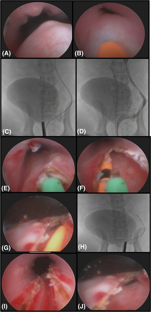
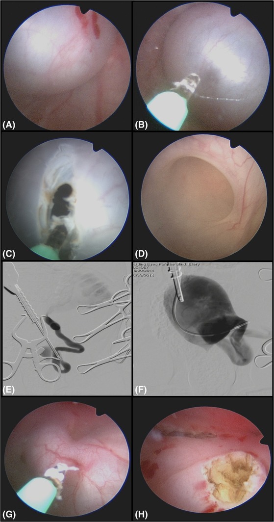
Ureteroceles are a rare condition in dogs in which conventional treatments can result in substantial morbidity. Cystoscopic and fluoroscopic-guided laser ablation (CLA) of ureteroceles can successfully relieve obstruction.
To describe the technique and outcomes of attempting CLA for treatment of ureteroceles in dogs.
Thirteen client-owned dogs that underwent CLA for treatment of ureteroceles.
Retrospective multicentered study. Medical records were reviewed in all dogs that underwent CLA for ureterocele(s). A laser was used to extend the opening of the ureteral orifice (UO) unless surgical conversion was necessary. Data collected included signalment, clinicopathologic data, imaging, procedural findings, complications, and short- and long-term outcome.
Thirteen dogs with 13 ureteroceles associated with 14 UOs resulting in ureteral obstruction were included. One ureterocele extended bilaterally. Treatment was initiated via retrograde cystoscopy (7 females), percutaneous perineal urethrocystoscopy (4 males), or percutaneous antegrade cystoscopy (2 males). Surgical conversion was necessary in 2 males. Ten of 14 (71%) UOs associated with the ureteroceles were ectopic. Thirteen of 14 had stenotic or imperforate UOs. No postoperative complications were noted. Preoperative incontinence or pollakiuria was present in 9 of 13 and 3 of 13 dogs and resolved in 8 of 9 and 3 of 3 dogs, respectively. Follow-up imaging showed resolution of all ureteroceles and improved ureteral/renal pelvic dilatation. Median follow-up time was 27 months (range, 3-96 months).
Cystoscopic-guided laser ablation was effective for the treatment of ureteroceles(s) in 11 of 13 dogs.
输尿管囊肿在犬类中是一种罕见病症,传统治疗可能导致严重的发病率。输尿管囊肿的膀胱镜和透视引导下激光消融(CLA)可成功缓解梗阻。
描述尝试采用CLA治疗犬输尿管囊肿的技术及结果。
13只接受CLA治疗输尿管囊肿的客户拥有的犬。
回顾性多中心研究。对所有接受CLA治疗输尿管囊肿的犬的病历进行回顾。除非需要手术转换,否则使用激光扩大输尿管口(UO)的开口。收集的数据包括信号、临床病理数据、影像学、手术结果、并发症以及短期和长期结果。
纳入13只患有与14个UO相关的13个输尿管囊肿并导致输尿管梗阻的犬。1个输尿管囊肿双侧延伸。治疗通过逆行膀胱镜检查(7只雌性)、经皮会阴尿道膀胱镜检查(4只雄性)或经皮顺行膀胱镜检查(2只雄性)开始。2只雄性需要手术转换。与输尿管囊肿相关的14个UO中有10个(71%)为异位。14个中有13个UO狭窄或无孔。未观察到术后并发症。13只犬中有9只术前存在尿失禁或尿频,13只中有3只存在尿频,分别在9只中的8只和3只中的3只中得到缓解。随访影像学显示所有输尿管囊肿均消退,输尿管/肾盂扩张改善。中位随访时间为27个月(范围3 - 96个月)。
膀胱镜引导下激光消融对13只犬中的11只输尿管囊肿治疗有效。