Mendoza-Azpur Gerardo, Olaechea Allinson, Padial-Molina Miguel, Gutiérrez-Garrido Lourdes, O'Valle Francisco, Mesa Francisco, Galindo-Moreno Pablo
Department of Periodontology, School of Dentistry, Cientifica del Sur University, 15067 Lima, Peru.
Department of Oral Surgery and Implant Dentistry, School of Dentistry, University of Granada, 18071 Granada, Spain.
J Clin Med. 2019 Feb 9;8(2):223. doi: 10.3390/jcm8020223.
The aim of this study was to examine the clinical and histological differences of using a combination of alloplastic beta triphasic calcium phosphate (β-TCP) and a cross-linked collagen membrane versus autologous platelet-rich fibrin (PRF-L) in ridge preservation after dental extraction.
Fifty-one patients were included in this observational case-series study. Dental extractions were performed, after which 25 patients were grafted with β-TCP and 26 with PRF-L. After four months of healing, clinical, radiological, histomorphometric and histological evaluations were performed.
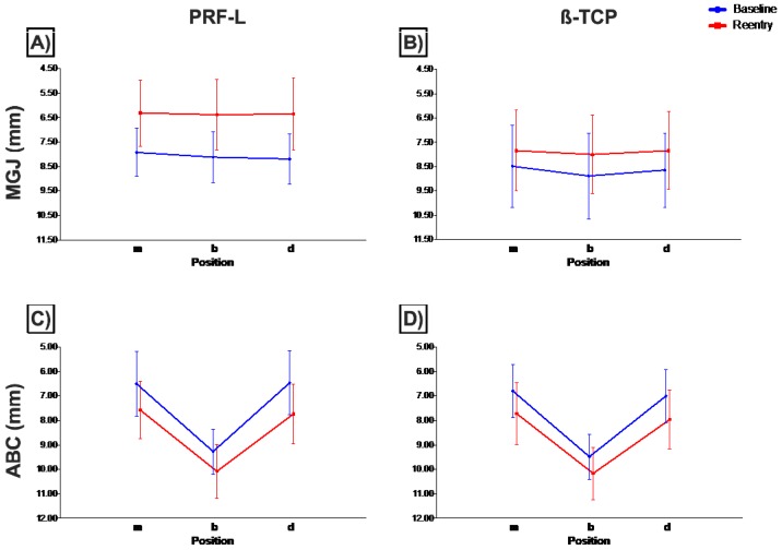
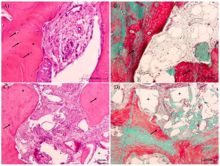
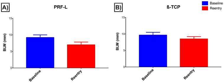
A significantly higher percentage of mineralized tissue was observed in samples from the PRF-L grafted areas. Cellularity was higher in PRF-L grafted areas (osteocytes in newly formed bone per mm² = 123.25 (5.12) vs. 84.02 (26.53) for PRF-L and β-TCP, respectively, = 0.01). However, sockets grafted with PRF-L showed a higher reduction in the bucco-lingual dimension after four months of healing (2.19 (0.80) vs. 1.16 (0.55) mm, < 0.001), as well as a higher alteration in the final position of the mid muco-gingival junction (1.73 (1.34) vs. 0.88 (0.88) mm, < 0.01).
PRF-L concentrate accelerates wound healing in post-extraction sockets in terms of new mineralized tissue component. However, the use of β-TCP biomaterial appears to be superior to maintain bucco-lingual volume and the final position of the muco-gingival junction.
本研究旨在探讨在拔牙后牙槽嵴保存中,使用异体β-三相磷酸钙(β-TCP)与交联胶原膜联合材料和自体富血小板纤维蛋白(PRF-L)的临床及组织学差异。
本观察性病例系列研究纳入了51例患者。进行拔牙后,25例患者植入β-TCP,26例患者植入PRF-L。愈合四个月后,进行临床、放射学、组织形态计量学和组织学评估。
在PRF-L植入区域的样本中观察到矿化组织的百分比显著更高。PRF-L植入区域的细胞密度更高(每平方毫米新形成骨中的骨细胞,PRF-L为123.25(5.12),β-TCP为84.02(26.53),P = 0.01)。然而,愈合四个月后,植入PRF-L的牙槽窝在颊舌径上的减小更大(2.19(0.80)对1.16(0.55)mm,P < 0.001),并且龈黏膜交界处最终位置的改变也更大(1.73(1.34)对0.88(0.88)mm,P < 0.01)。
就新的矿化组织成分而言,PRF-L浓缩物可加速拔牙后牙槽窝的伤口愈合。然而,使用β-TCP生物材料在维持颊舌体积和龈黏膜交界处的最终位置方面似乎更具优势。