• 文献检索
  • 文档翻译
  • 深度研究
  • 学术资讯
  • Suppr Zotero 插件Zotero 插件
  • 邀请有礼
  • 套餐&价格
  • 历史记录
应用&插件
Suppr Zotero 插件Zotero 插件浏览器插件Mac 客户端Windows 客户端微信小程序
定价
高级版会员购买积分包购买API积分包
服务
文献检索文档翻译深度研究API 文档MCP 服务
关于我们
关于 Suppr公司介绍联系我们用户协议隐私条款
关注我们

Suppr 超能文献

核心技术专利:CN118964589B侵权必究
粤ICP备2023148730 号-1Suppr @ 2026

文献检索

告别复杂PubMed语法,用中文像聊天一样搜索,搜遍4000万医学文献。AI智能推荐,让科研检索更轻松。

立即免费搜索

文件翻译

保留排版,准确专业,支持PDF/Word/PPT等文件格式,支持 12+语言互译。

免费翻译文档

深度研究

AI帮你快速写综述,25分钟生成高质量综述,智能提取关键信息,辅助科研写作。

立即免费体验

应用二次谐波显微镜对交联后角膜胶原分布的定量分析。

Quantitative Analysis of the Corneal Collagen Distribution after Cross-Linking with Second Harmonic Microscopy.

机构信息

Laboratorio de Óptica, Instituto Universitario de Investigación en Óptica y Nanofísica, Universidad de Murcia, Campus de Espinardo (Ed. 34), 30100 Murcia, Spain.

Dpto. Biología Celular, Histología y Farmacología, Facultad de Medicina, Universidad de Valladolid, 47005 Valladolid, Spain.

出版信息

Biomed Res Int. 2019 Jan 10;2019:3860498. doi: 10.1155/2019/3860498. eCollection 2019.

DOI:10.1155/2019/3860498
PMID:30756083
原文链接:https://pmc.ncbi.nlm.nih.gov/articles/PMC6348900/
Abstract

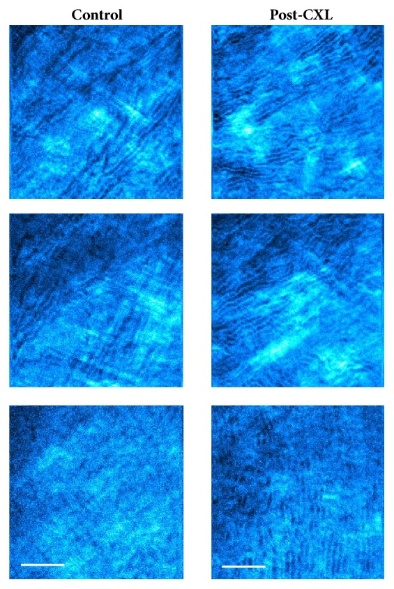
Corneal cross-linking (CXL) is a surgical procedure able to modify corneal biomechanics and stabilize keratoconus progression. Although it is known that CXL produces changes in corneal collagen distribution, these are still a topic of discussion. Here we quantitatively compare the corneal stroma architecture between two animal models four weeks after conventional CXL treatment, with second harmonic generation (SHG) imaging microscopy and the structure tensor (ST). The healing stage and the stroma recovery were also analyzed by means of histological sections. Results show that the CXL effects depend on the initial arrangement of the corneal collagen. While the treatment increases the order in corneas with a low level of initial organization, corneas presenting a fairly regular pattern are hardly affected. Histological samples showed active keratocytes in anterior and middle stroma, what means that the recovery is still in progress. The combination of SHG imaging and the ST is able to objectively discriminate the changes suffered by the collagen arrangement after the CXL treatment, whose effectiveness depends on the initial organization of the collagen fibers within the corneal stroma.

摘要

角膜交联术(CXL)是一种能够改变角膜生物力学并稳定圆锥角膜进展的手术。尽管已知 CXL 会改变角膜胶原的分布,但这仍然是一个讨论的话题。在这里,我们使用二次谐波产生(SHG)成像显微镜和结构张量(ST),定量比较了两种动物模型在传统 CXL 治疗后四周的角膜基质结构。还通过组织学切片分析了愈合阶段和基质恢复情况。结果表明,CXL 效应取决于角膜胶原的初始排列。虽然该治疗会增加初始组织程度较低的角膜的有序性,但对排列较规则的角膜几乎没有影响。组织学样本显示前部和中部基质中有活跃的角膜细胞,这意味着恢复仍在进行中。SHG 成像和 ST 的结合能够客观地区分 CXL 治疗后胶原排列所经历的变化,其有效性取决于角膜基质中胶原纤维的初始组织程度。

https://cdn.ncbi.nlm.nih.gov/pmc/blobs/0706/6348900/7c14ede0e031/BMRI2019-3860498.011.jpg
https://cdn.ncbi.nlm.nih.gov/pmc/blobs/0706/6348900/fcb7d655db9b/BMRI2019-3860498.001.jpg
https://cdn.ncbi.nlm.nih.gov/pmc/blobs/0706/6348900/d455f326cf55/BMRI2019-3860498.002.jpg
https://cdn.ncbi.nlm.nih.gov/pmc/blobs/0706/6348900/7745daa5bce5/BMRI2019-3860498.003.jpg
https://cdn.ncbi.nlm.nih.gov/pmc/blobs/0706/6348900/d4db08629e78/BMRI2019-3860498.004.jpg
https://cdn.ncbi.nlm.nih.gov/pmc/blobs/0706/6348900/bd8ea4667d29/BMRI2019-3860498.005.jpg
https://cdn.ncbi.nlm.nih.gov/pmc/blobs/0706/6348900/76433307a23a/BMRI2019-3860498.006.jpg
https://cdn.ncbi.nlm.nih.gov/pmc/blobs/0706/6348900/30196d47423c/BMRI2019-3860498.007.jpg
https://cdn.ncbi.nlm.nih.gov/pmc/blobs/0706/6348900/b8f66bdedc29/BMRI2019-3860498.008.jpg
https://cdn.ncbi.nlm.nih.gov/pmc/blobs/0706/6348900/5e773dfe3ff1/BMRI2019-3860498.009.jpg
https://cdn.ncbi.nlm.nih.gov/pmc/blobs/0706/6348900/7083ffaddc30/BMRI2019-3860498.010.jpg
https://cdn.ncbi.nlm.nih.gov/pmc/blobs/0706/6348900/7c14ede0e031/BMRI2019-3860498.011.jpg

相似文献

1
Quantitative Analysis of the Corneal Collagen Distribution after Cross-Linking with Second Harmonic Microscopy.应用二次谐波显微镜对交联后角膜胶原分布的定量分析。
Biomed Res Int. 2019 Jan 10;2019:3860498. doi: 10.1155/2019/3860498. eCollection 2019.
2
Morphological and immunohistochemical changes after corneal cross-linking.角膜交联术后的形态和免疫组织化学变化。
Cornea. 2013 Feb;32(2):111-7. doi: 10.1097/ICO.0b013e31824d701b.
3
Corneal confocal microscopy following conventional, transepithelial, and accelerated corneal collagen cross-linking procedures for keratoconus.角膜共焦显微镜检查在常规、经上皮和加速角膜胶原交联程序治疗圆锥角膜中的应用。
J Refract Surg. 2012 Nov;28(11):769-76. doi: 10.3928/1081597X-20121016-01.
4
In vivo confocal microscopy analyses of corneal microstructural changes in a prospective study of collagen cross-linking in keratoconus.一项前瞻性研究中角膜交联术治疗圆锥角膜的角膜微观结构变化的共焦显微镜分析。
Ophthalmology. 2014 Feb;121(2):469-74. doi: 10.1016/j.ophtha.2013.09.014. Epub 2013 Oct 30.
5
Corneal Nerve Regeneration After Collagen Cross-Linking Treatment of Keratoconus: A 5-Year Longitudinal Study.圆锥角膜胶原交联治疗后的角膜神经再生:一项5年纵向研究。
JAMA Ophthalmol. 2016 Jan;134(1):70-8. doi: 10.1001/jamaophthalmol.2015.4518.
6
Matrix-Based Regenerating Agent for Corneal Wound Healing After Collagen Cross-Linking.基于基质的胶原蛋白交联后角膜伤口愈合再生剂
Cornea. 2016 Dec;35(12):1638-1643. doi: 10.1097/ICO.0000000000001047.
7
Multiphoton microscopy of ex vivo corneas after collagen cross-linking.胶原交联术后离体角膜的多光子显微镜检查。
Invest Ophthalmol Vis Sci. 2011 Jul 18;52(8):5325-31. doi: 10.1167/iovs.11-7184.
8
Morphological and functional correlations in riboflavin UV A corneal collagen cross-linking for keratoconus.核黄素 UVA 角膜胶原交联术治疗圆锥角膜的形态与功能相关性。
Acta Ophthalmol. 2012 May;90(3):259-65. doi: 10.1111/j.1755-3768.2010.01890.x. Epub 2010 Apr 23.
9
In Vivo Confocal Microscopy After Contact Lens-Assisted Corneal Collagen Cross-linking for Thin Keratoconic Corneas.软性角膜接触镜辅助角膜胶原交联治疗薄圆锥角膜后的体内共聚焦显微镜检查
J Refract Surg. 2016 May 1;32(5):326-31. doi: 10.3928/1081597X-20160225-04.
10
Transepithelial riboflavin/ultraviolet. a corneal cross-linking in keratoconus: morphologic studies on human corneas.经上皮角膜胶原交联术在圆锥角膜中的应用:对人眼角膜的形态学研究。
Am J Ophthalmol. 2013 Nov;156(5):874-884.e1. doi: 10.1016/j.ajo.2013.06.025. Epub 2013 Aug 20.

引用本文的文献

1
Quantitative Characterization of Corneal Collagen Architecture Using Intensity Gradient Modeling and Gaussian PDF Fitting.使用强度梯度建模和高斯概率密度函数拟合对角膜胶原结构进行定量表征。
Diagnostics (Basel). 2025 Jul 8;15(14):1738. doi: 10.3390/diagnostics15141738.
2
On the quantitative analysis of lamellar collagen arrangement with second-harmonic generation imaging.基于二次谐波成像的层状胶原排列定量分析
Biomed Opt Express. 2024 Mar 29;15(4):2666-2680. doi: 10.1364/BOE.516817. eCollection 2024 Apr 1.
3
Characterization of pathological stomach tissue using polarization-sensitive second harmonic generation microscopy.

本文引用的文献

1
Rose Bengal and Green Light Versus Riboflavin-UVA Cross-Linking: Corneal Wound Repair Response.玫瑰红 Bengal 和绿光与核黄素-UVA 交联:角膜伤口修复反应。
Invest Ophthalmol Vis Sci. 2018 Oct 1;59(12):4821-4830. doi: 10.1167/iovs.18-24881.
2
Conventional and transepithelial corneal cross-linking for patients with keratoconus.传统经上皮角膜交联术与角膜交联术治疗圆锥角膜。
PLoS One. 2018 Apr 5;13(4):e0195105. doi: 10.1371/journal.pone.0195105. eCollection 2018.
3
High-resolution, label-free two-photon imaging of diseased human corneas.
使用偏振敏感二次谐波产生显微镜对病理性胃组织进行表征。
Biomed Opt Express. 2023 Sep 22;14(10):5376-5391. doi: 10.1364/BOE.500335. eCollection 2023 Oct 1.
4
A review of the epithelial and stromal effects of corneal collagen crosslinking.角膜胶原交联的上皮和基质效应评价。
Ocul Surf. 2023 Oct;30:150-159. doi: 10.1016/j.jtos.2023.09.003. Epub 2023 Sep 6.
5
Two-Photon Imaging for Non-Invasive Corneal Examination.双光子成像技术在非侵入性角膜检查中的应用。
Sensors (Basel). 2022 Dec 11;22(24):9699. doi: 10.3390/s22249699.
6
Examining lysyl oxidase-like modulation of collagen architecture in 3D spheroid models of idiopathic pulmonary fibrosis via second-harmonic generation microscopy.通过二次谐波产生显微镜检查特发性肺纤维化 3D 球体模型中赖氨酰氧化酶样对胶原结构的调节。
J Biomed Opt. 2021 Jun;26(6). doi: 10.1117/1.JBO.26.6.066501.
7
Corneal Optical Coherence Tomography Speckle in Crosslinked and Untreated Rabbit Eyes in Response to Elevated Intraocular Pressure.交联和未处理兔眼对眼内压升高的角膜光学相干断层扫描散斑反应。
Transl Vis Sci Technol. 2021 Apr 29;10(5):2. doi: 10.1167/tvst.10.5.2.
8
Assessment of the corneal collagen organization after chemical burn using second harmonic generation microscopy.使用二次谐波产生显微镜评估化学烧伤后的角膜胶原组织。
Biomed Opt Express. 2021 Jan 11;12(2):756-765. doi: 10.1364/BOE.412819. eCollection 2021 Feb 1.
9
Assessment of progressive alterations in collagen organization in the postoperative conjunctiva by multiphoton microscopy.通过多光子显微镜评估术后结膜中胶原蛋白组织的渐进性改变。
Biomed Opt Express. 2020 Oct 19;11(11):6495-6515. doi: 10.1364/BOE.403555. eCollection 2020 Nov 1.
10
Nonlinear optical crosslinking (NLO CXL) for correcting refractive errors.非线性光学交联(NLO CXL)矫正屈光不正。
Exp Eye Res. 2020 Oct;199:108199. doi: 10.1016/j.exer.2020.108199. Epub 2020 Aug 23.
高分辨率、无标记的人眼角膜疾病双光子成像。
J Biomed Opt. 2018 Mar;23(3):1-8. doi: 10.1117/1.JBO.23.3.036002.
4
Quantization of collagen organization in the stroma with a new order coefficient.用新的有序系数对基质中胶原蛋白组织进行量化。
Biomed Opt Express. 2017 Dec 8;9(1):173-189. doi: 10.1364/BOE.9.000173. eCollection 2018 Jan 1.
5
Assessment of Human Corneas Prior to Transplantation Using High-Resolution Two-Photon Imaging.高分辨率双光子成像在移植前对人眼角膜的评估。
Invest Ophthalmol Vis Sci. 2018 Jan 1;59(1):176-184. doi: 10.1167/iovs.17-22002.
6
Corneal Wound Repair After Rose Bengal and Green Light Crosslinking: Clinical and Histologic Study.孟加拉玫瑰红和绿光交联后的角膜伤口修复:临床与组织学研究
Invest Ophthalmol Vis Sci. 2017 Jul 1;58(9):3471-3480. doi: 10.1167/iovs.16-21365.
7
Multimodal imaging quality control of epithelia regenerated with cultured human donor corneal limbal epithelial stem cells.培养的人供体角膜缘上皮干细胞再生的上皮的多模态成像质量控制。
Sci Rep. 2017 Jul 11;7(1):5154. doi: 10.1038/s41598-017-05486-8.
8
Randomized Controlled Trial Comparing Transepithelial Corneal Cross-linking Using Iontophoresis with the Dresden Protocol in Progressive Keratoconus.离子导入诱导交联与 Dresden 方案治疗进展性圆锥角膜的随机对照临床试验
Ophthalmology. 2017 Jun;124(6):804-812. doi: 10.1016/j.ophtha.2017.01.040. Epub 2017 Mar 7.
9
Improved quantification of collagen anisotropy with polarization-resolved second harmonic generation microscopy.利用偏振分辨二次谐波产生显微镜改进胶原蛋白各向异性的定量分析。
J Biophotonics. 2017 Sep;10(9):1171-1179. doi: 10.1002/jbio.201600197. Epub 2016 Oct 24.
10
Three-dimensional mapping of the orientation of collagen corneal lamellae in healthy and keratoconic human corneas using SHG microscopy.使用二次谐波产生显微镜对健康和圆锥角膜人眼角膜中胶原角膜板层的方向进行三维映射。
J Biophotonics. 2017 Jan;10(1):75-83. doi: 10.1002/jbio.201600122. Epub 2016 Jul 29.