• 文献检索
  • 文档翻译
  • 深度研究
  • 学术资讯
  • Suppr Zotero 插件Zotero 插件
  • 邀请有礼
  • 套餐&价格
  • 历史记录
应用&插件
Suppr Zotero 插件Zotero 插件浏览器插件Mac 客户端Windows 客户端微信小程序
定价
高级版会员购买积分包购买API积分包
服务
文献检索文档翻译深度研究API 文档MCP 服务
关于我们
关于 Suppr公司介绍联系我们用户协议隐私条款
关注我们

Suppr 超能文献

核心技术专利:CN118964589B侵权必究
粤ICP备2023148730 号-1Suppr @ 2026

文献检索

告别复杂PubMed语法,用中文像聊天一样搜索,搜遍4000万医学文献。AI智能推荐,让科研检索更轻松。

立即免费搜索

文件翻译

保留排版,准确专业,支持PDF/Word/PPT等文件格式,支持 12+语言互译。

免费翻译文档

深度研究

AI帮你快速写综述,25分钟生成高质量综述,智能提取关键信息,辅助科研写作。

立即免费体验

使用拉曼光谱技术对乳腺癌与良性肿瘤进行鉴别诊断。

Discrimination of breast cancer from benign tumours using Raman spectroscopy.

机构信息

Centre for Radiation and Environmental Science, FOCAS Research Institute, Technological University Dublin, Dublin, Ireland.

Department of Chemistry and Earth Sciences, Qatar University, Doha, Qatar.

出版信息

PLoS One. 2019 Feb 14;14(2):e0212376. doi: 10.1371/journal.pone.0212376. eCollection 2019.

DOI:10.1371/journal.pone.0212376
PMID:30763392
原文链接:https://pmc.ncbi.nlm.nih.gov/articles/PMC6375635/
Abstract

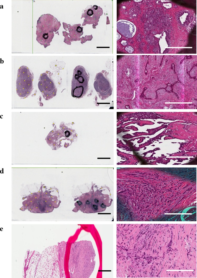
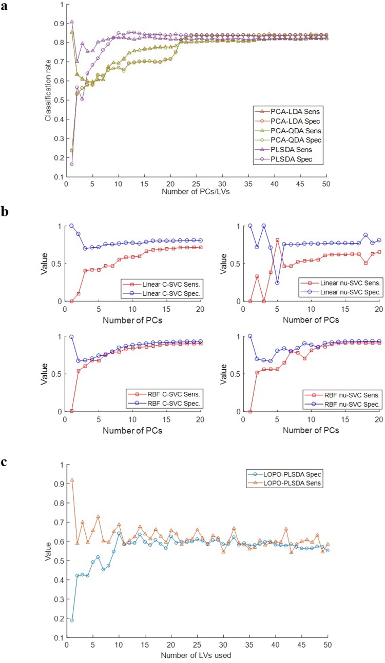
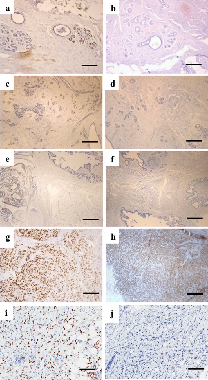
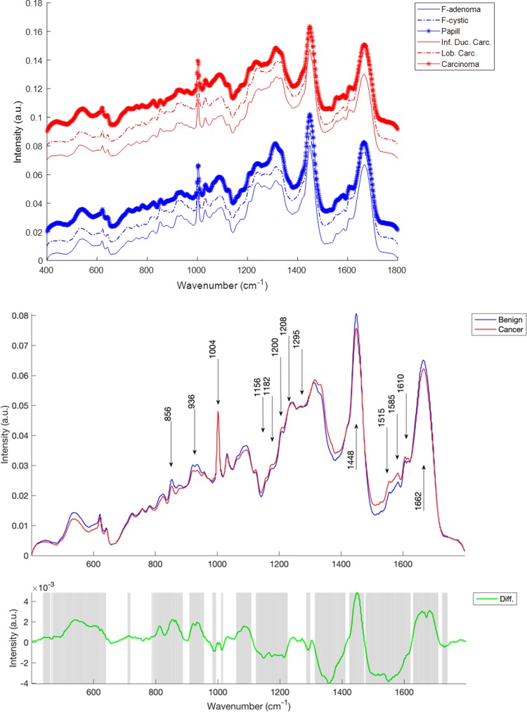
Breast cancer is the most common cancer among women worldwide, with an estimated 1.7 million cases and 522,000 deaths in 2012. Breast cancer is diagnosed by histopathological examination of breast biopsy material but this is subjective and relies on morphological changes in the tissue. Raman spectroscopy uses incident radiation to induce vibrations in the molecules of a sample and the scattered radiation can be used to characterise the sample. This technique is rapid and non-destructive and is sensitive to subtle biochemical changes occurring at the molecular level. This allows spectral variations corresponding to disease onset to be detected. The aim of this work was to use Raman spectroscopy to discriminate between benign lesions (fibrocystic, fibroadenoma, intraductal papilloma) and cancer (invasive ductal carcinoma and lobular carcinoma) using formalin fixed paraffin preserved (FFPP) tissue. Haematoxylin and Eosin stained sections from the patient biopsies were marked by a pathologist. Raman maps were recorded from parallel unstained tissue sections. Immunohistochemical staining for estrogen receptor (ER) and human epidermal growth factor receptor 2 (HER2/neu) was performed on a further set of parallel sections. Both benign and cancer cases were positive for ER while only the cancer cases were positive for HER2. Significant spectral differences were observed between the benign and cancer cases and the benign cases could be differentiated from the cancer cases with good sensitivity and specificity. This study has shown the potential of Raman spectroscopy as an aid to histopathological diagnosis of breast cancer, in particular in the discrimination between benign and malignant tumours.

摘要

乳腺癌是全世界女性最常见的癌症,据估计,2012 年全球有 170 万例乳腺癌新发病例和 52.2 万例死亡病例。乳腺癌的诊断是通过对乳腺活检材料的组织病理学检查来实现的,但这种方法具有主观性,并且依赖于组织的形态变化。拉曼光谱技术利用入射辐射诱导样品分子的振动,散射辐射可用于对样品进行特征描述。这种技术快速、无损,对分子水平上发生的微妙生化变化非常敏感。这使得可以检测到与疾病发生相对应的光谱变化。本工作的目的是使用拉曼光谱技术,通过对福尔马林固定石蜡包埋(FFPP)组织的良性病变(纤维囊性、纤维腺瘤、导管内乳头状瘤)和癌症(浸润性导管癌和小叶癌)进行区分。从患者活检中提取的经苏木精和伊红染色的切片由病理学家进行标记。从平行的未染色组织切片中记录拉曼图谱。对另一组平行切片进行雌激素受体(ER)和人表皮生长因子受体 2(HER2/neu)的免疫组织化学染色。良性和癌症病例均对 ER 呈阳性,而只有癌症病例对 HER2 呈阳性。良性和癌症病例之间观察到明显的光谱差异,并且可以通过高灵敏度和特异性将良性病例与癌症病例区分开来。本研究表明拉曼光谱技术具有作为辅助组织病理学诊断乳腺癌的潜力,特别是在良性和恶性肿瘤的鉴别方面。

https://cdn.ncbi.nlm.nih.gov/pmc/blobs/1cfb/6375635/a4487f17c434/pone.0212376.g004.jpg
https://cdn.ncbi.nlm.nih.gov/pmc/blobs/1cfb/6375635/45ffb32bca71/pone.0212376.g001.jpg
https://cdn.ncbi.nlm.nih.gov/pmc/blobs/1cfb/6375635/e0de898a3a04/pone.0212376.g002.jpg
https://cdn.ncbi.nlm.nih.gov/pmc/blobs/1cfb/6375635/298fc542889a/pone.0212376.g003.jpg
https://cdn.ncbi.nlm.nih.gov/pmc/blobs/1cfb/6375635/a4487f17c434/pone.0212376.g004.jpg

相似文献

1
Discrimination of breast cancer from benign tumours using Raman spectroscopy.使用拉曼光谱技术对乳腺癌与良性肿瘤进行鉴别诊断。
PLoS One. 2019 Feb 14;14(2):e0212376. doi: 10.1371/journal.pone.0212376. eCollection 2019.
2
Identification of Molecular Basis for Objective Discrimination of Breast Cancer Cells (MCF-7) from Normal Human Mammary Epithelial Cells by Raman Microspectroscopy and Multivariate Curve Resolution Analysis.拉曼光谱和多变量曲线分辨分析鉴定客观区分乳腺癌细胞(MCF-7)和正常人类乳腺上皮细胞的分子基础。
Int J Mol Sci. 2021 Jan 14;22(2):800. doi: 10.3390/ijms22020800.
3
The use of Au@SiO2 shell-isolated nanoparticle-enhanced Raman spectroscopy for human breast cancer detection.金@二氧化硅壳层隔离纳米粒子增强拉曼光谱在人类乳腺癌检测中的应用。
Anal Bioanal Chem. 2014 Sep;406(22):5425-32. doi: 10.1007/s00216-014-7967-5. Epub 2014 Jun 24.
4
Raman spectra exploring breast tissues: comparison of principal component analysis and support vector machine-recursive feature elimination.拉曼光谱探测乳腺组织:主成分分析和支持向量机-递归特征消除的比较。
Med Phys. 2013 Jun;40(6):063501. doi: 10.1118/1.4804054.
5
Differences and Relationships Between Normal and Atypical Ductal Hyperplasia, Ductal Carcinoma In Situ, and Invasive Ductal Carcinoma Tissues in the Breast Based on Raman Spectroscopy.基于拉曼光谱的乳腺正常与非典型导管增生、导管原位癌及浸润性导管癌组织之间的差异与关系
Appl Spectrosc. 2017 Feb;71(2):300-307. doi: 10.1177/0003702816681009. Epub 2016 Dec 20.
6
[Expression of fatty acid synthase and its association with HER2 in invasive ductal carcinoma of breast].[脂肪酸合酶在乳腺浸润性导管癌中的表达及其与HER2的关系]
Zhonghua Bing Li Xue Za Zhi. 2013 Apr;42(4):257-61. doi: 10.3760/cma.j.issn.0529-5807.2013.04.010.
7
Expression of c-kit in common benign and malignant breast lesions.c-kit在常见乳腺良恶性病变中的表达
Tumori. 2010 Nov-Dec;96(6):978-84.
8
Breast Tumor Analysis Using Shifted-Excitation Raman Difference Spectroscopy (SERDS).使用位移激发拉曼差分光谱(SERDS)进行乳腺肿瘤分析。
Technol Cancer Res Treat. 2018 Jan 1;17:1533033818782532. doi: 10.1177/1533033818782532.
9
Estrogen receptor, progesterone receptor, and human epidermal growth factor receptor 2 expression in breast cancer FNA cell blocks and paired histologic specimens: A large retrospective study.雌激素受体、孕激素受体及人表皮生长因子受体2在乳腺癌细针穿刺抽吸细胞块及配对组织学标本中的表达:一项大型回顾性研究。
Cancer Cytopathol. 2016 Nov;124(11):828-835. doi: 10.1002/cncy.21745. Epub 2016 Jun 17.
10
A Molecular Typing Method for Invasive Breast Cancer by Serum Raman Spectroscopy.血清拉曼光谱法用于浸润性乳腺癌的分子分型。
Clin Breast Cancer. 2024 Jun;24(4):376-383. doi: 10.1016/j.clbc.2024.02.008. Epub 2024 Feb 16.

引用本文的文献

1
Raman difference spectroscopy and U-Net convolutional neural network for molecular analysis of cutaneous neurofibroma.拉曼差分光谱和 U-Net 卷积神经网络在皮肤神经纤维瘤的分子分析中的应用。
PLoS One. 2024 Apr 11;19(4):e0302017. doi: 10.1371/journal.pone.0302017. eCollection 2024.
2
Use of Raman spectroscopy to study rat lung tissues for distinguishing asphyxia from sudden cardiac death.利用拉曼光谱研究大鼠肺组织以区分窒息与心源性猝死。
RSC Adv. 2024 Feb 14;14(8):5665-5674. doi: 10.1039/d3ra07684a. eCollection 2024 Feb 7.
3
Cellular dynamics as a marker of normal-to-cancer transition in human cells.

本文引用的文献

1
Raman spectroscopy for cancer detection and cancer surgery guidance: translation to the clinics.拉曼光谱学在癌症检测和癌症手术指导中的应用:向临床的转化。
Analyst. 2017 Aug 21;142(17):3025-3047. doi: 10.1039/c7an00957g.
2
A review of Raman spectroscopy advances with an emphasis on clinical translation challenges in oncology.拉曼光谱学进展综述:着重于肿瘤学临床转化面临的挑战
Phys Med Biol. 2016 Dec 7;61(23):R370-R400. doi: 10.1088/0031-9155/61/23/R370. Epub 2016 Nov 2.
3
Application of Raman Spectroscopy and Infrared Spectroscopy in the Identification of Breast Cancer.
细胞动力学作为人细胞正常向癌变转化的标志物。
Sci Rep. 2023 Nov 29;13(1):21079. doi: 10.1038/s41598-023-47649-w.
4
Molecular Fingerprint Detection Using Raman and Infrared Spectroscopy Technologies for Cancer Detection: A Progress Review.利用拉曼和红外光谱技术进行分子指纹检测在癌症检测中的应用:进展综述。
Biosensors (Basel). 2023 May 18;13(5):557. doi: 10.3390/bios13050557.
5
A Machine Learning Framework for Detecting COVID-19 Infection Using Surface-Enhanced Raman Scattering.基于表面增强拉曼散射的 COVID-19 感染检测机器学习框架。
Biosensors (Basel). 2022 Aug 2;12(8):589. doi: 10.3390/bios12080589.
6
Vibrational Biospectroscopy: An Alternative Approach to Endometrial Cancer Diagnosis and Screening.振动生物光谱学:子宫内膜癌诊断和筛查的另一种方法。
Int J Mol Sci. 2022 Apr 27;23(9):4859. doi: 10.3390/ijms23094859.
7
A New Look into Cancer-A Review on the Contribution of Vibrational Spectroscopy on Early Diagnosis and Surgery Guidance.癌症新视角——振动光谱在早期诊断和手术指导中的作用综述
Cancers (Basel). 2021 Oct 24;13(21):5336. doi: 10.3390/cancers13215336.
8
Surface-Enhanced Raman Spectroscopy Characterization of Breast Cell Phenotypes: Effect of Nanoparticle Geometry.乳腺细胞表型的表面增强拉曼光谱表征:纳米颗粒几何形状的影响
ACS Appl Nano Mater. 2019 Nov 22;2(11):6960-6970. doi: 10.1021/acsanm.9b01436. Epub 2019 Oct 20.
9
Raman Spectral Signatures of Serum-Derived Extracellular Vesicle-Enriched Isolates May Support the Diagnosis of CNS Tumors.血清来源的富含细胞外囊泡的分离物的拉曼光谱特征可能有助于中枢神经系统肿瘤的诊断。
Cancers (Basel). 2021 Mar 19;13(6):1407. doi: 10.3390/cancers13061407.
10
Diagnostic accuracy of Raman spectroscopy for prostate cancer: a systematic review and meta-analysis.拉曼光谱对前列腺癌的诊断准确性:一项系统评价和荟萃分析。
Transl Androl Urol. 2021 Feb;10(2):574-583. doi: 10.21037/tau-20-924.
拉曼光谱和红外光谱在乳腺癌鉴别中的应用。
Appl Spectrosc. 2016 Feb;70(2):251-63. doi: 10.1177/0003702815620127.
4
New look inside human breast ducts with Raman imaging. Raman candidates as diagnostic markers for breast cancer prognosis: Mammaglobin, palmitic acid and sphingomyelin.利用拉曼成像技术对人类乳腺导管进行新观察。拉曼光谱可作为乳腺癌预后诊断标志物:乳腺珠蛋白、棕榈酸和鞘磷脂。
Anal Chim Acta. 2016 Feb 25;909:91-100. doi: 10.1016/j.aca.2015.12.038. Epub 2016 Jan 4.
5
Raman spectroscopy for medical diagnostics--From in-vitro biofluid assays to in-vivo cancer detection.拉曼光谱在医学诊断中的应用——从体外生物流体分析到体内癌症检测。
Adv Drug Deliv Rev. 2015 Jul 15;89:121-34. doi: 10.1016/j.addr.2015.03.009. Epub 2015 Mar 22.
6
Raman microspectroscopy of noncancerous and cancerous human breast tissues. Identification and phase transitions of linoleic and oleic acids by Raman low-temperature studies.人乳腺非癌组织和癌组织的拉曼显微光谱。通过拉曼低温研究鉴定亚油酸和油酸及其相变。
Analyst. 2015 Apr 7;140(7):2134-43. doi: 10.1039/c4an01877j.
7
The lipid-reactive oxygen species phenotype of breast cancer. Raman spectroscopy and mapping, PCA and PLSDA for invasive ductal carcinoma and invasive lobular carcinoma. Molecular tumorigenic mechanisms beyond Warburg effect.乳腺癌的脂质活性氧表型。浸润性导管癌和浸润性小叶癌的拉曼光谱及成像、主成分分析和偏最小二乘判别分析。超越瓦伯格效应的分子致瘤机制。
Analyst. 2015 Apr 7;140(7):2121-33. doi: 10.1039/c4an01876a.
8
Towards intra-operative diagnosis of tumours during breast conserving surgery by selective-sampling Raman micro-spectroscopy.通过选择性采样拉曼显微光谱术实现保乳手术中肿瘤的术中诊断。
Phys Med Biol. 2014 Oct 21;59(20):6141-52. doi: 10.1088/0031-9155/59/20/6141. Epub 2014 Sep 25.
9
Cancer incidence and mortality worldwide: sources, methods and major patterns in GLOBOCAN 2012.全球癌症发病与死亡:GLOBOCAN 2012 数据源、方法与主要模式。
Int J Cancer. 2015 Mar 1;136(5):E359-86. doi: 10.1002/ijc.29210. Epub 2014 Oct 9.
10
Breast cancer and melanoma cell line identification by FTIR imaging after formalin-fixation and paraffin-embedding.傅里叶变换红外成像技术在福尔马林固定石蜡包埋后对乳腺癌和黑色素瘤细胞系的鉴定。
Analyst. 2013 Jul 21;138(14):4083-91. doi: 10.1039/c3an00246b.