Gebrekidan Medhanie Tesfay, Erber Ramona, Hartmann Arndt, Fasching Peter A, Emons Julius, Beckmann Mathias W, Braeuer Andreas
1 Lehrstuhl für Technische Thermodynamik, Friedrich-Alexander-Universität (FAU), Erlangen-Nürnberg, Germany.
2 Erlangen Graduate School in Advanced Optical Technologies (SAOT), Friedrich-Alexander-Universität (FAU), Erlangen-Nürnberg, Germany.
Technol Cancer Res Treat. 2018 Jan 1;17:1533033818782532. doi: 10.1177/1533033818782532.
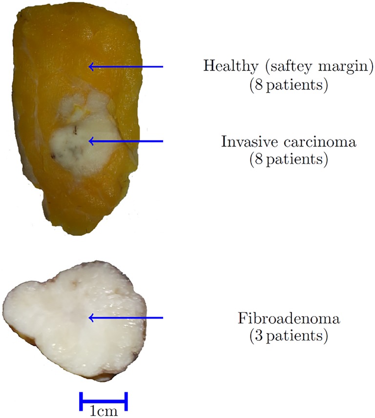
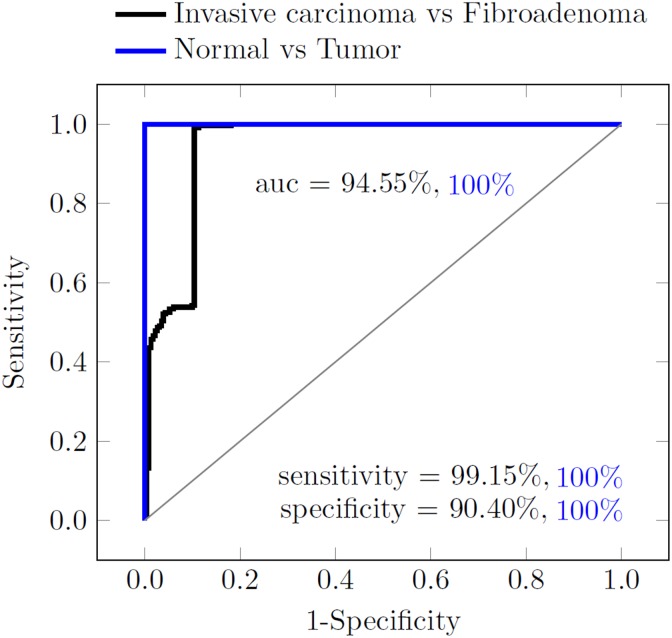
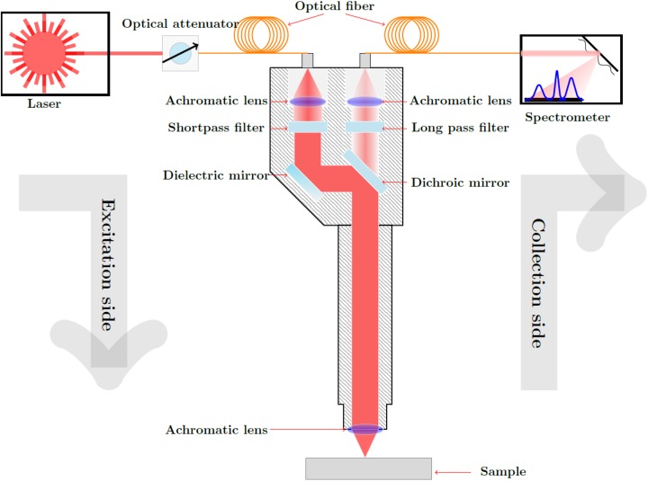
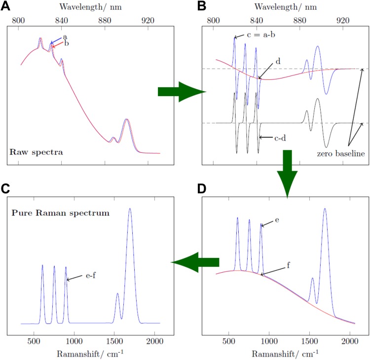
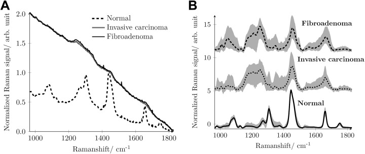
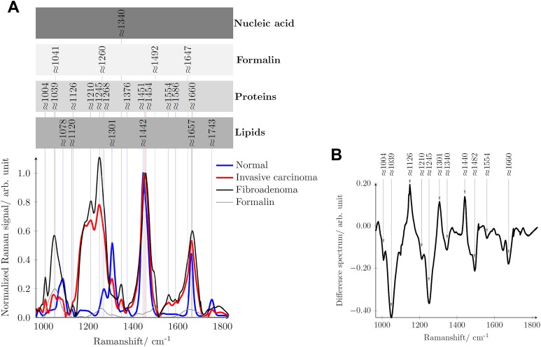
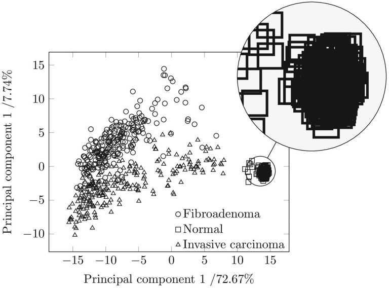
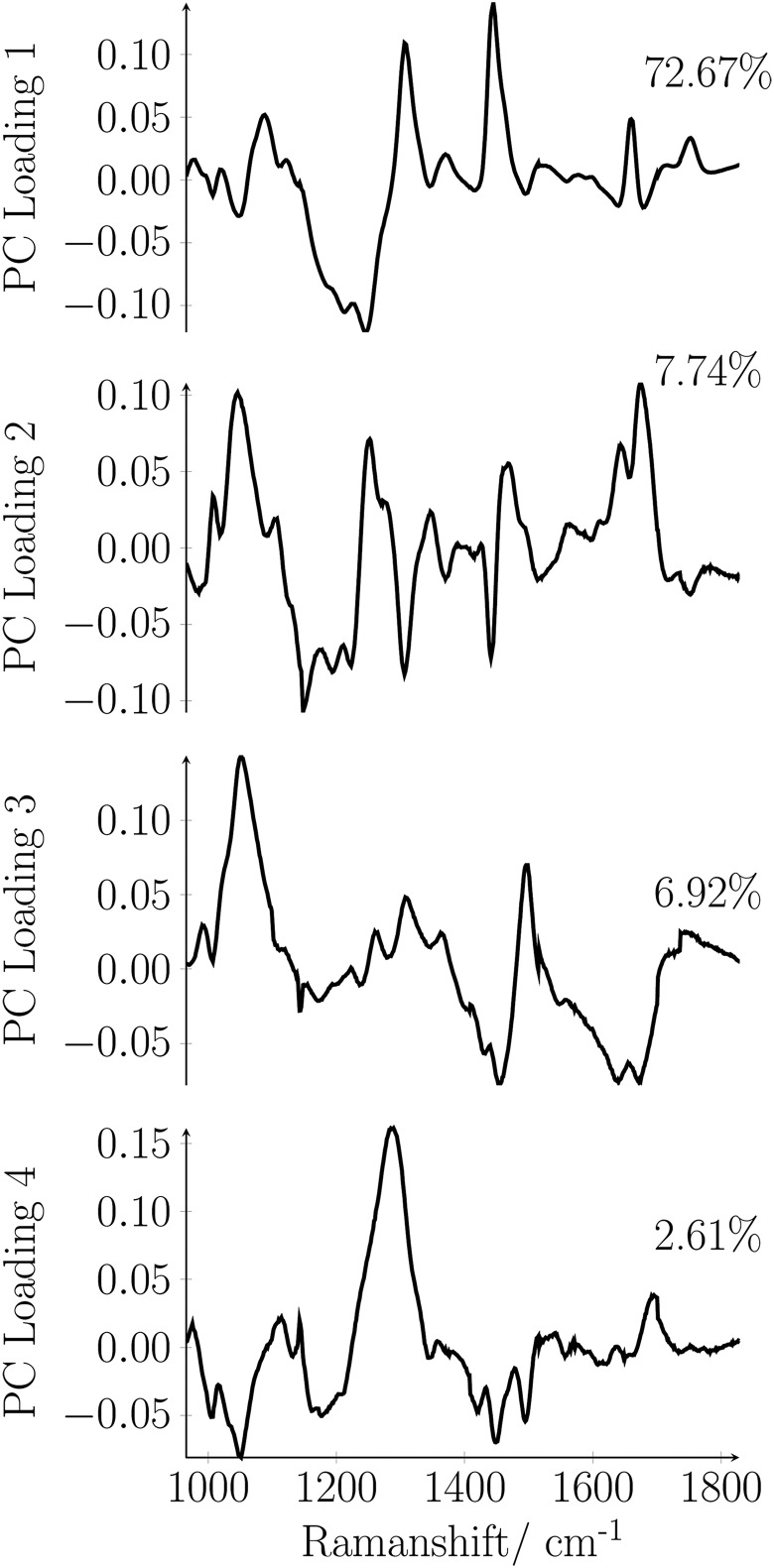
We used a shifted-excitation Raman difference spectroscopy method for the ex vivo classification of resected and formalin-fixed breast tissue samples as normal (healthy) tissue, fibroadenoma, or invasive carcinoma. We analyzed 8 tissue samples containing invasive carcinoma that were surrounded by normal tissue and 3 tissue samples with fibroadenoma only. We made various measurement sites on various tissue samples, in total 240 measurements for each type of tissue. Although the acquired raw spectra contain enough information to clearly differentiate between normal and tumor (fibroadenoma and invasive carcinoma) tissue, the differentiation between fibroadenoma and invasive carcinoma was possible only after the shifted-excitation Raman difference spectroscopy isolation of pure Raman spectra from the heavily fluorescence interfered raw spectra. We used 784 and 785 nm as excitation wavelengths for the shifted-excitation Raman difference spectroscopy method. The differences in the obtained pure Raman spectra are assigned to the different chemical compositions of normal breast tissue, fibroadenoma, and invasive breast carcinoma. Principal component analysis and linear discriminant analysis showed excellent classification results in the Raman shift range between 1000 and 1800 cm. Invasive breast carcinoma was identified with 99.15% sensitivity, and the absence of invasive carcinoma was identified with 90.40% specificity. Tumor tissue in tumor-containing tissue was identified with 100% sensitivity, and the absence of tumor in no-tumor containing tissue was identified with 100% specificity. As gold standard for the determination of the sensitivity and the specificity, we considered the conventional histopathological classification. In summary, shifted-excitation Raman difference spectroscopy could be potentially very useful to support histopathological diagnosis in breast pathology.
我们采用位移激发拉曼差分光谱法对切除的、经福尔马林固定的乳腺组织样本进行离体分类,区分正常(健康)组织、纤维腺瘤或浸润性癌。我们分析了8个含有被正常组织包围的浸润性癌的组织样本以及3个仅含纤维腺瘤的组织样本。我们在各种组织样本上设置了不同的测量位点,每种组织类型总共进行了240次测量。尽管采集到的原始光谱包含足够的信息以清晰区分正常组织和肿瘤(纤维腺瘤和浸润性癌)组织,但只有在从严重受荧光干扰的原始光谱中通过位移激发拉曼差分光谱法分离出纯拉曼光谱后,才有可能区分纤维腺瘤和浸润性癌。我们将784和785 nm用作位移激发拉曼差分光谱法的激发波长。所获得的纯拉曼光谱中的差异归因于正常乳腺组织、纤维腺瘤和浸润性乳腺癌的不同化学成分。主成分分析和线性判别分析在1000至1800 cm的拉曼位移范围内显示出优异的分类结果。浸润性乳腺癌的识别灵敏度为99.15%,未发现浸润性癌的识别特异性为90.40%。含肿瘤组织中的肿瘤组织识别灵敏度为100%,不含肿瘤组织中未发现肿瘤的识别特异性为100%。作为确定灵敏度和特异性的金标准,我们采用了传统的组织病理学分类。总之,位移激发拉曼差分光谱法在乳腺病理学中可能对支持组织病理学诊断非常有用。