• 文献检索
  • 文档翻译
  • 深度研究
  • 学术资讯
  • Suppr Zotero 插件Zotero 插件
  • 邀请有礼
  • 套餐&价格
  • 历史记录
应用&插件
Suppr Zotero 插件Zotero 插件浏览器插件Mac 客户端Windows 客户端微信小程序
定价
高级版会员购买积分包购买API积分包
服务
文献检索文档翻译深度研究API 文档MCP 服务
关于我们
关于 Suppr公司介绍联系我们用户协议隐私条款
关注我们

Suppr 超能文献

核心技术专利:CN118964589B侵权必究
粤ICP备2023148730 号-1Suppr @ 2026

文献检索

告别复杂PubMed语法,用中文像聊天一样搜索,搜遍4000万医学文献。AI智能推荐,让科研检索更轻松。

立即免费搜索

文件翻译

保留排版,准确专业,支持PDF/Word/PPT等文件格式,支持 12+语言互译。

免费翻译文档

深度研究

AI帮你快速写综述,25分钟生成高质量综述,智能提取关键信息,辅助科研写作。

立即免费体验

常规流式细胞术检测尿液中外泌体的高灵敏度免疫捕获。

High sensitivity detection of extracellular vesicles immune-captured from urine by conventional flow cytometry.

机构信息

Department of Immunology and Oncology, National Centre for Biotechnology, CNB-CSIC, Madrid, Spain.

Department of Molecular Biology, UAM, Centro de Biología Molecular Severo Ochoa (CBM-SO), Instituto de Investigación Sanitaria Princesa (IIS-IP), Madrid, Spain.

出版信息

Sci Rep. 2019 Feb 14;9(1):2042. doi: 10.1038/s41598-019-38516-8.

DOI:10.1038/s41598-019-38516-8
PMID:30765839
原文链接:https://pmc.ncbi.nlm.nih.gov/articles/PMC6376115/
Abstract

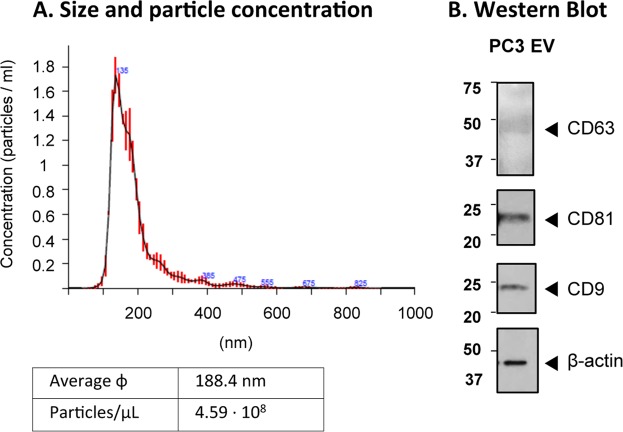
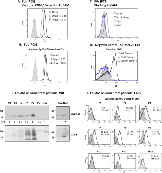
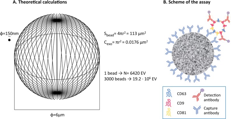
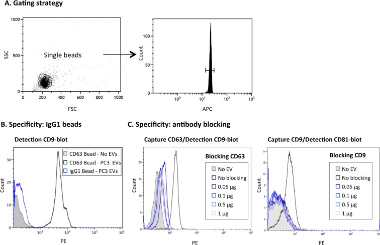
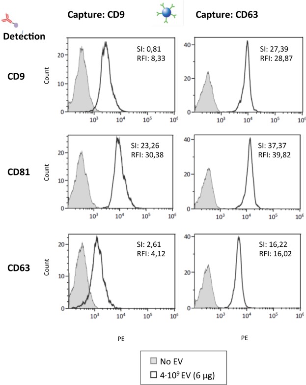
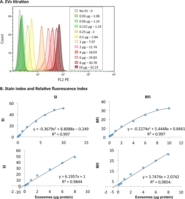
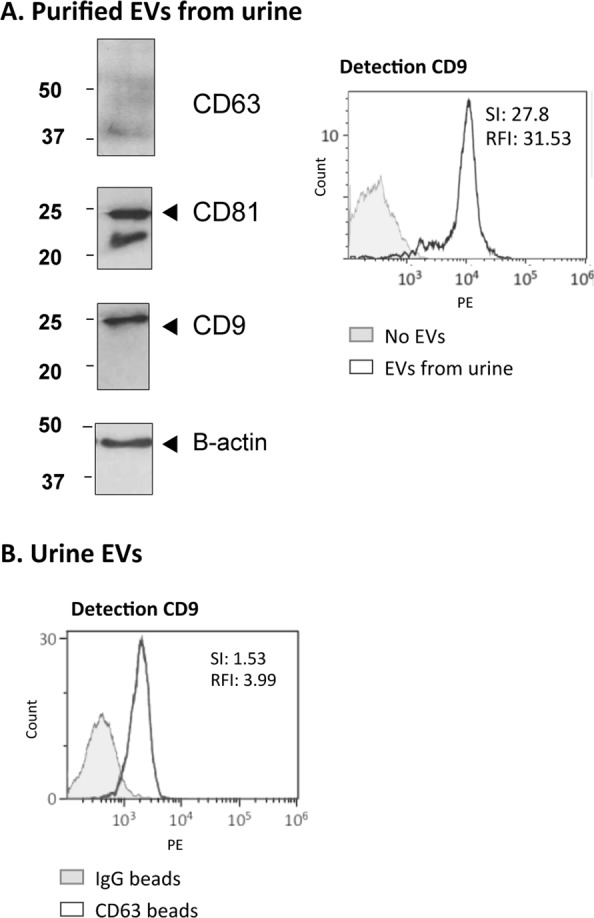
Extracellular vesicles (EVs) provide an invaluable tool to analyse physiological processes because they transport, in biological fluids, biomolecules secreted from diverse tissues of an individual. EV biomarker detection requires highly sensitive techniques able to identify individual molecules. However, the lack of widespread, affordable methodologies for high-throughput EV analyses means that studies on biomarkers have not been done in large patient cohorts. To develop tools for EV analysis in biological samples, we evaluated here the critical parameters to optimise an assay based on immunocapture of EVs followed by flow cytometry. We describe a straightforward method for EV detection using general EV markers like the tetraspanins CD9, CD63 and CD81, that allowed highly sensitive detection of urinary EVs without prior enrichment. In proof-of-concept experiments, an epithelial marker enriched in carcinoma cells, EpCAM, was identified in EVs from cell lines and directly in urine samples. However, whereas EVs isolated from 5-10 ml of urine were required for western blot detection of EpCAM, only 500 μl of urine were sufficient to visualise EpCAM expression by flow cytometry. This method has the potential to allow any laboratory with access to conventional flow cytometry to identify surface markers on EVs, even non-abundant proteins, using minimally processed biological samples.

摘要

细胞外囊泡 (EVs) 为分析生理过程提供了非常有价值的工具,因为它们在生物体液中运输个体不同组织分泌的生物分子。EV 生物标志物检测需要高度敏感的技术,以识别单个分子。然而,缺乏广泛且经济实惠的高通量 EV 分析方法,意味着在大型患者队列中尚未进行生物标志物研究。为了开发生物样本中 EV 分析的工具,我们在这里评估了优化基于 EV 免疫捕获的流式细胞术的分析方法的关键参数。我们描述了一种使用一般 EV 标志物(如四跨膜蛋白 CD9、CD63 和 CD81)检测 EV 的简单方法,该方法无需预先富集即可高度敏感地检测尿液中的 EV。在概念验证实验中,在细胞系和直接在尿液样本中,上皮细胞标志物 EpCAM 被鉴定为存在于 EV 中。然而,尽管用于 Western blot 检测 EpCAM 需要分离来自 5-10ml 尿液的 EV,但仅需 500μl 尿液即可通过流式细胞术观察到 EpCAM 的表达。这种方法有可能使任何能够获得常规流式细胞术的实验室,使用最小处理的生物样本,识别 EV 上的表面标志物,甚至是非丰富的蛋白质。

https://cdn.ncbi.nlm.nih.gov/pmc/blobs/7462/6376115/4e65605c2301/41598_2019_38516_Fig7_HTML.jpg
https://cdn.ncbi.nlm.nih.gov/pmc/blobs/7462/6376115/bb22fb15f774/41598_2019_38516_Fig1_HTML.jpg
https://cdn.ncbi.nlm.nih.gov/pmc/blobs/7462/6376115/dcdf72de92ec/41598_2019_38516_Fig2_HTML.jpg
https://cdn.ncbi.nlm.nih.gov/pmc/blobs/7462/6376115/ad71da26d45b/41598_2019_38516_Fig3_HTML.jpg
https://cdn.ncbi.nlm.nih.gov/pmc/blobs/7462/6376115/d1942e53142c/41598_2019_38516_Fig4_HTML.jpg
https://cdn.ncbi.nlm.nih.gov/pmc/blobs/7462/6376115/6395059ce855/41598_2019_38516_Fig5_HTML.jpg
https://cdn.ncbi.nlm.nih.gov/pmc/blobs/7462/6376115/9080b9fced75/41598_2019_38516_Fig6_HTML.jpg
https://cdn.ncbi.nlm.nih.gov/pmc/blobs/7462/6376115/4e65605c2301/41598_2019_38516_Fig7_HTML.jpg

相似文献

1
High sensitivity detection of extracellular vesicles immune-captured from urine by conventional flow cytometry.常规流式细胞术检测尿液中外泌体的高灵敏度免疫捕获。
Sci Rep. 2019 Feb 14;9(1):2042. doi: 10.1038/s41598-019-38516-8.
2
Nanoscale flow cytometry to distinguish subpopulations of prostate extracellular vesicles in patient plasma.纳米流式细胞术可区分患者血浆中前列腺细胞外囊泡的亚群。
Prostate. 2019 May;79(6):592-603. doi: 10.1002/pros.23764. Epub 2019 Jan 24.
3
Surface protein profiling of milk and serum extracellular vesicles unveils body fluid-specific signatures.对奶和血清细胞外囊泡的表面蛋白进行分析,揭示了体液特异性特征。
Sci Rep. 2023 May 30;13(1):8758. doi: 10.1038/s41598-023-35799-w.
4
Large Extracellular Vesicles Can be Characterised by Multiplex Labelling Using Imaging Flow Cytometry.大型细胞外囊泡可通过成像流式细胞术进行多重标记来表征。
Int J Mol Sci. 2020 Nov 18;21(22):8723. doi: 10.3390/ijms21228723.
5
Assessment of technical and clinical utility of a bead-based flow cytometry platform for multiparametric phenotyping of CNS-derived extracellular vesicles.评估基于珠粒的流式细胞仪平台在中枢神经系统来源细胞外囊泡的多参数表型分析中的技术和临床实用性。
Cell Commun Signal. 2023 Oct 6;21(1):276. doi: 10.1186/s12964-023-01308-9.
6
Single Extracellular Vesicle Analysis Performed by Imaging Flow Cytometry and Nanoparticle Tracking Analysis Evaluate the Accuracy of Urinary Extracellular Vesicle Preparation Techniques Differently.成像流式细胞术和纳米颗粒跟踪分析对单个细胞外囊泡的分析评估了尿液细胞外囊泡准备技术的准确性。
Int J Mol Sci. 2021 Nov 18;22(22):12436. doi: 10.3390/ijms222212436.
7
An Immunocapture-Based Assay for Detecting Multiple Antigens in Melanoma-Derived Extracellular Vesicles.基于免疫捕获的方法检测黑色素瘤衍生细胞外囊泡中的多种抗原。
Methods Mol Biol. 2021;2265:323-344. doi: 10.1007/978-1-0716-1205-7_24.
8
Extracellular vesicles isolated from porcine seminal plasma exhibit different tetraspanin expression profiles.从猪精液中分离出的细胞外囊泡表现出不同的四跨膜蛋白表达谱。
Sci Rep. 2019 Aug 9;9(1):11584. doi: 10.1038/s41598-019-48095-3.
9
Differential proteomics argues against a general role for CD9, CD81 or CD63 in the sorting of proteins into extracellular vesicles.差异蛋白质组学表明 CD9、CD81 或 CD63 一般不会参与将蛋白质分选到细胞外囊泡中。
J Extracell Vesicles. 2023 Aug;12(8):e12352. doi: 10.1002/jev2.12352.
10
Comparison of Generic Fluorescent Markers for Detection of Extracellular Vesicles by Flow Cytometry.通用荧光标记物在流式细胞术检测细胞外囊泡中的比较。
Clin Chem. 2018 Apr;64(4):680-689. doi: 10.1373/clinchem.2017.278978. Epub 2018 Feb 16.

引用本文的文献

1
Labeling, isolation and characterization of cell-type-specific exosomes derived from mouse skin tissue.源自小鼠皮肤组织的细胞类型特异性外泌体的标记、分离与鉴定
Nat Protoc. 2025 Sep 12. doi: 10.1038/s41596-025-01238-5.
2
Proteomic profiling of extracellular vesicles distinguishes prostate cancer molecular subtypes.细胞外囊泡的蛋白质组学分析可区分前列腺癌分子亚型。
bioRxiv. 2025 Aug 2:2025.07.31.667906. doi: 10.1101/2025.07.31.667906.
3
The Roles of Exosomes in Anti-Cancer Drugs.外泌体在抗癌药物中的作用。

本文引用的文献

1
Immunoassays for scarce tumour-antigens in exosomes: detection of the human NKG2D-Ligand, MICA, in tetraspanin-containing nanovesicles from melanoma.外泌体中稀有肿瘤抗原的免疫分析:在含有四跨膜蛋白的纳米囊泡中检测黑色素瘤的人类 NKG2D-Ligand(MICA)。
J Nanobiotechnology. 2018 May 2;16(1):47. doi: 10.1186/s12951-018-0372-z.
2
Immunoaffinity-based isolation of melanoma cell-derived exosomes from plasma of patients with melanoma.基于免疫亲和从黑色素瘤患者血浆中分离黑色素瘤细胞衍生的外泌体。
J Extracell Vesicles. 2018 Feb 15;7(1):1435138. doi: 10.1080/20013078.2018.1435138. eCollection 2018.
3
Impaired NK cell recognition of vemurafenib-treated melanoma cells is overcome by simultaneous application of histone deacetylase inhibitors.
Cancer Med. 2025 May;14(9):e70897. doi: 10.1002/cam4.70897.
4
Affinity-Based Isolation and One-Pot Analysis of Extracellular Vesicles from Biofluids Using Phase Separated Zwitterionic Coacervates.基于亲和力的生物流体中细胞外囊泡的分离及使用相分离两性离子凝聚层的一锅法分析
Adv Sci (Weinh). 2025 May;12(20):e2411653. doi: 10.1002/advs.202411653. Epub 2025 Apr 15.
5
Utilizing Two Electrokinetic Techniques on a Single Device for Detection of Extracellular Vesicle-Associated Protease Activity.在单个设备上利用两种电动技术检测细胞外囊泡相关蛋白酶活性。
Small. 2025 Jun;21(24):e2404457. doi: 10.1002/smll.202404457. Epub 2025 Mar 26.
6
Mechanistic insight into human milk extracellular vesicle-intestinal barrier interactions.人乳细胞外囊泡与肠道屏障相互作用的机制洞察
J Extracell Biol. 2025 Jan 9;4(1):e70032. doi: 10.1002/jex2.70032. eCollection 2025 Jan.
7
Squeezable Hydrogel Microparticles for Single Extracellular Vesicle Protein Profiling.用于单细胞外囊泡蛋白质谱分析的可挤压水凝胶微粒
Small. 2025 Jan;21(1):e2407809. doi: 10.1002/smll.202407809. Epub 2024 Oct 29.
8
Investigating miR-6880-5p in extracellular vesicle from plasma as a prognostic biomarker in endocrine therapy-treated castration-resistant prostate cancer.探讨血浆外泌体中的 miR-6880-5p 作为内分泌治疗后去势抵抗性前列腺癌的预后生物标志物。
BMC Cancer. 2024 Jul 29;24(1):909. doi: 10.1186/s12885-024-12460-x.
9
Optimization of extracellular vesicle isolation and their separation from lipoproteins by size exclusion chromatography.通过尺寸排阻色谱法优化细胞外囊泡的分离及其与脂蛋白的分离。
J Extracell Biol. 2023 Jul 8;2(7):e100. doi: 10.1002/jex2.100. eCollection 2023 Jul.
10
Comparing digital detection platforms in high sensitivity immune-phenotyping of extracellular vesicles.比较数字检测平台在细胞外囊泡高灵敏度免疫表型分析中的应用
J Extracell Biol. 2022 Aug 3;1(8):e53. doi: 10.1002/jex2.53. eCollection 2022 Aug.
同时应用组蛋白去乙酰化酶抑制剂可克服维莫非尼治疗的黑色素瘤细胞自然杀伤细胞识别受损的问题。
Oncoimmunology. 2017 Nov 6;7(2):e1392426. doi: 10.1080/2162402X.2017.1392426. eCollection 2018.
4
Protein Profiling and Sizing of Extracellular Vesicles from Colorectal Cancer Patients via Flow Cytometry.通过流式细胞术对结直肠癌患者的细胞外囊泡进行蛋白质分析和大小分析。
ACS Nano. 2018 Jan 23;12(1):671-680. doi: 10.1021/acsnano.7b07782. Epub 2018 Jan 8.
5
A bead-assisted flow cytometry method for the semi-quantitative analysis of Extracellular Vesicles.一种基于珠子的流式细胞术方法,用于外泌体的半定量分析。
Sci Rep. 2017 Sep 12;7(1):11271. doi: 10.1038/s41598-017-11249-2.
6
Labeling Extracellular Vesicles for Nanoscale Flow Cytometry.标记细胞外囊泡用于纳米尺度流式细胞术。
Sci Rep. 2017 May 12;7(1):1878. doi: 10.1038/s41598-017-01731-2.
7
Exosomes: From Garbage Bins to Promising Therapeutic Targets.外泌体:从垃圾桶到有前景的治疗靶点
Int J Mol Sci. 2017 Mar 2;18(3):538. doi: 10.3390/ijms18030538.
8
Flow Cytometric Analysis of Extracellular Vesicles.细胞外囊泡的流式细胞术分析
Methods Mol Biol. 2017;1545:215-225. doi: 10.1007/978-1-4939-6728-5_16.
9
Isolation of Exosomes and Microvesicles from Cell Culture Systems to Study Prion Transmission.从细胞培养系统中分离外泌体和微囊泡以研究朊病毒传播
Methods Mol Biol. 2017;1545:153-176. doi: 10.1007/978-1-4939-6728-5_11.
10
Size-Exclusion Chromatography-based isolation minimally alters Extracellular Vesicles' characteristics compared to precipitating agents.基于尺寸排阻色谱法的分离与沉淀剂相比,最小程度地改变了细胞外囊泡的特征。
Sci Rep. 2016 Sep 19;6:33641. doi: 10.1038/srep33641.