Department of Laboratory Medicine, Division of Pathology F46, Karolinska Institutet, Karolinska University Hospital Huddinge, SE-141 86, Stockholm, Sweden.
Department of Clinical Pathology/Cytology, Karolinska University Hospital, Stockholm, SE-141 86, Sweden.
Sci Rep. 2019 Feb 14;9(1):2133. doi: 10.1038/s41598-019-38603-w.
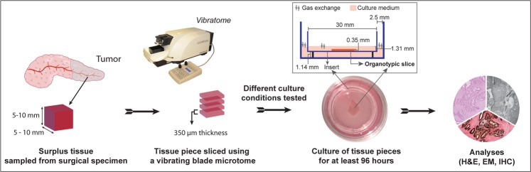
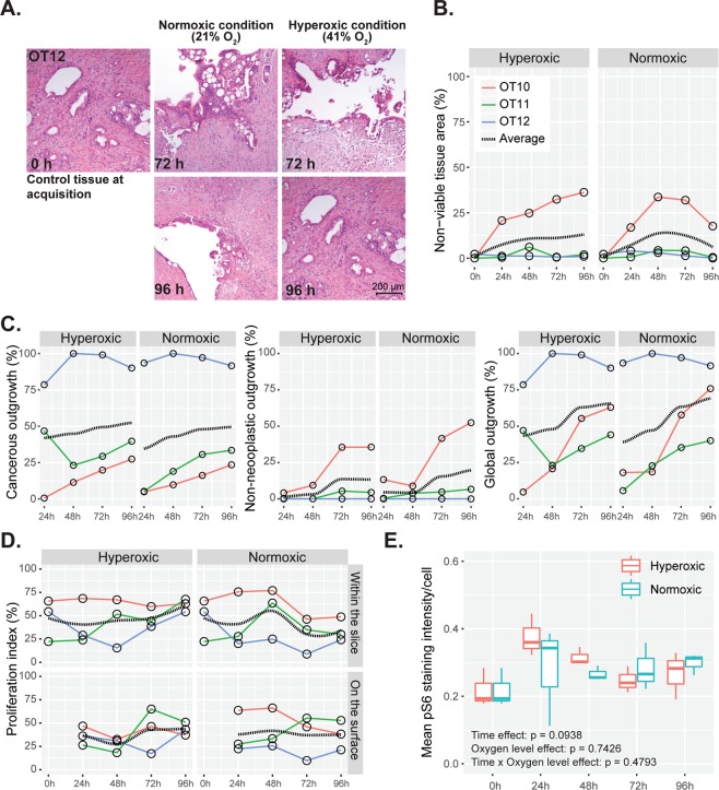
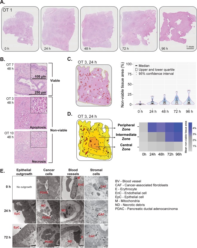
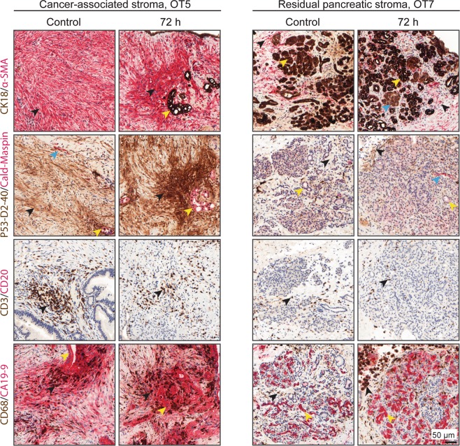
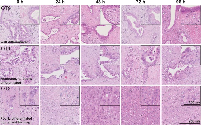
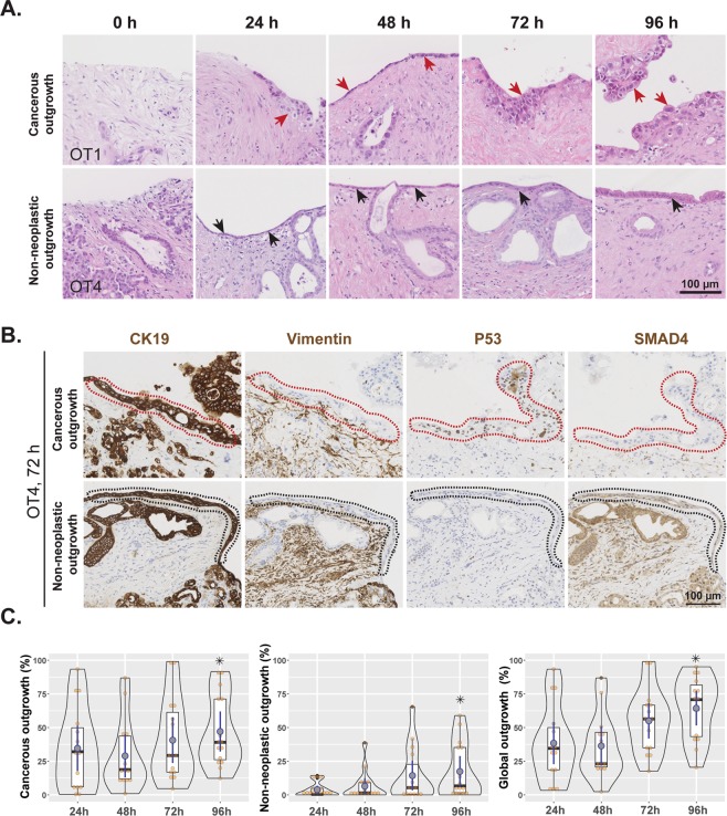
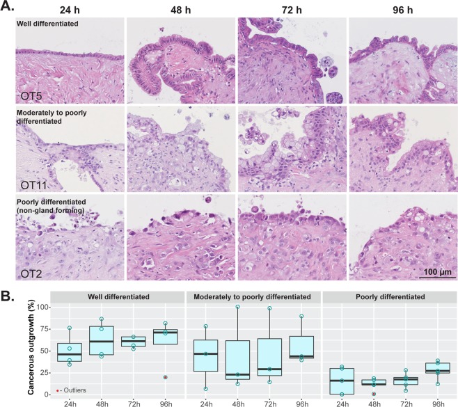
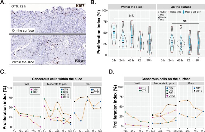
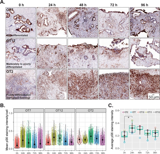
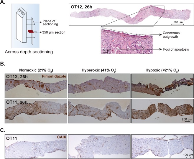
Pancreatic ductal adenocarcinoma (PDAC) has a poor prognosis, which is mainly due to late diagnosis and profound resistance to treatment. The latter is to a large extent attributed to the tumor stroma that is exceedingly prominent in PDAC and engages in complex interactions with the cancer cells. Hence, relevant preclinical models of PDAC should also include the tumor stroma. We herein describe the establishment and functional validation of an ex vivo organotypic culture of human PDAC that is based on precision-cut tissue slices from surgical specimens and reproducibly recapitulates the complex cellular and acellular composition of PDAC, including its microenvironment. The cancer cells, tumor microenvironment and interspersed remnants of nonneoplastic pancreas contained in these 350 µm thick slices maintained their structural integrity, phenotypic characteristics and functional activity when in culture for at least 4 days. In particular, tumor cell proliferation persisted and the grade of differentiation and morphological phenotype remained unaltered. Cultured tissue slices were metabolically active and responsive to rapamycin, an mTOR inhibitor. This culture system is to date the closest surrogate to the parent carcinoma and harbors great potential as a drug sensitivity testing system for the personalized treatment of PDAC.
胰腺导管腺癌(PDAC)预后较差,这主要是由于诊断较晚和对治疗的深度抵抗。后者在很大程度上归因于 PDAC 中非常突出的肿瘤基质,并与癌细胞进行复杂的相互作用。因此,相关的 PDAC 临床前模型也应包括肿瘤基质。我们在此描述了一种基于手术标本的精确切割组织切片的人 PDAC 离体器官型培养的建立和功能验证,该培养能够重现 PDAC 的复杂细胞和无细胞组成,包括其微环境。当在培养至少 4 天时,这些 350μm 厚切片中的癌细胞、肿瘤微环境和穿插的非肿瘤胰腺残余物保持其结构完整性、表型特征和功能活性。特别是,肿瘤细胞增殖持续存在,分化程度和形态表型保持不变。培养的组织切片具有代谢活性,并对雷帕霉素(mTOR 抑制剂)有反应。到目前为止,这种培养系统是与母体癌最接近的替代物,并且具有作为 PDAC 个体化治疗的药物敏感性测试系统的巨大潜力。