Chao Shih-Chun, Yang Shang-Jung, Chen Hung-Chi, Sun Chi-Chin, Liu Chin-Hsin, Lee Chia-Yi
Department of Ophthalmology, Show Chwan Memorial Hospital, Changhua, Taiwan.
Department of Electrical and Computer Engineering, National Chiao Tung University, Hsinchu, Taiwan.
J Ophthalmol. 2019 Jan 15;2019:7419470. doi: 10.1155/2019/7419470. eCollection 2019.
To evaluate early macular circulation in open-angle glaucoma (OAG), normal-tension glaucoma (NTG), ocular hypertension (OHT), and healthy subjects via optical coherence tomography angiography (OCTA).
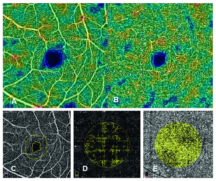
A retrospective cross-sectional study was conducted. Medical records were reviewed, and the patients who received OCTA examinations were divided into the OAG, NTG, OHT, and normal groups. The ophthalmic data including best-corrected visual acuity, spherical equivalent, intraocular pressure, central corneal thickness, central foveal thickness, visual field deviation, retinal nerve fiber layers thickness, and ganglion cell complex thickness were obtained from medical documents. For the macular area, the superficial vessel density (VD), deep VD, foveal avascular zone (FAZ), flow area of the outer retina, and flow area of the choriocapillaris were measured via OCTA and analyzed using the default vascular density analysis program in the same OCTA device.
A total of 70 eyes from 70 patients were analyzed in the current study. Significant differences in the intraocular pressure, central corneal thickness, visual field deviation, retinal fiber layer thickness, and ganglion cell complex thickness were observed in the patients in the glaucoma group at their last visits. The OAG and NTG groups evinced a lower superficial VD than did the control group, while the NTG group had a lower deep VD than the control group. The NTG group also had a larger FAZ than did the OHT group. The flow area of the outer retina in the OAG group was low relative to those of the OHT and control groups. No difference in choriocapillaris perfusion was observed among the groups.
The OAG and NTG patients demonstrated impaired vasculature before significant disease development could be observed. Furthermore, the differences in macular circulation may be associated with differences in the courses of disease between the glaucoma and OHT patients.
通过光学相干断层扫描血管造影(OCTA)评估开角型青光眼(OAG)、正常眼压性青光眼(NTG)、高眼压症(OHT)患者及健康受试者的早期黄斑循环。
进行一项回顾性横断面研究。查阅病历,将接受OCTA检查的患者分为OAG组、NTG组、OHT组和正常组。从病历中获取眼科数据,包括最佳矫正视力、等效球镜度、眼压、中央角膜厚度、中央凹厚度、视野偏差、视网膜神经纤维层厚度和神经节细胞复合体厚度。对于黄斑区,通过OCTA测量浅表血管密度(VD)、深层VD、黄斑无血管区(FAZ)、外层视网膜血流面积和脉络膜毛细血管血流面积,并使用同一OCTA设备中的默认血管密度分析程序进行分析。
本研究共分析了70例患者的70只眼。青光眼组患者末次就诊时,眼压、中央角膜厚度、视野偏差、视网膜纤维层厚度和神经节细胞复合体厚度存在显著差异。OAG组和NTG组的浅表VD低于对照组,而NTG组的深层VD低于对照组。NTG组的FAZ也大于OHT组。OAG组外层视网膜的血流面积相对于OHT组和对照组较低。各组之间未观察到脉络膜毛细血管灌注差异。
OAG和NTG患者在观察到明显疾病发展之前就已出现血管系统受损。此外,黄斑循环的差异可能与青光眼和OHT患者疾病进程的差异有关。