• 文献检索
  • 文档翻译
  • 深度研究
  • 学术资讯
  • Suppr Zotero 插件Zotero 插件
  • 邀请有礼
  • 套餐&价格
  • 历史记录
应用&插件
Suppr Zotero 插件Zotero 插件浏览器插件Mac 客户端Windows 客户端微信小程序
定价
高级版会员购买积分包购买API积分包
服务
文献检索文档翻译深度研究API 文档MCP 服务
关于我们
关于 Suppr公司介绍联系我们用户协议隐私条款
关注我们

Suppr 超能文献

核心技术专利:CN118964589B侵权必究
粤ICP备2023148730 号-1Suppr @ 2026

文献检索

告别复杂PubMed语法,用中文像聊天一样搜索,搜遍4000万医学文献。AI智能推荐,让科研检索更轻松。

立即免费搜索

文件翻译

保留排版,准确专业,支持PDF/Word/PPT等文件格式,支持 12+语言互译。

免费翻译文档

深度研究

AI帮你快速写综述,25分钟生成高质量综述,智能提取关键信息,辅助科研写作。

立即免费体验

正常眼压性青光眼中通过光学相干断层扫描血管造影术评估视野与视乳头周围血管密度的相关性

Correlation of Visual Field With Peripapillary Vessel Density Through Optical Coherence Tomography Angiography in Normal-Tension Glaucoma.

作者信息

Lin Yun-Hsuan, Huang Shih-Ming, Yeung Ling, Ku Wan-Chen, Chen Henry Shen-Lih, Lai Chi-Chun, Chuang Lan-Hsin

机构信息

Department of Electro-Optical Engineering, National Taipei University of Technology, Taipei, Taiwan.

Department of Ophthalmology, Chang Gung Memorial Hospital, Keelung, Taiwan.

出版信息

Transl Vis Sci Technol. 2020 Dec 17;9(13):26. doi: 10.1167/tvst.9.13.26. eCollection 2020 Dec.

DOI:10.1167/tvst.9.13.26
PMID:33364080
原文链接:https://pmc.ncbi.nlm.nih.gov/articles/PMC7746955/
Abstract

PURPOSE

To investigate the retinal vessel density (VD) in healthy and normal-tension glaucoma (NTG) eyes through optical coherence tomography angiography (OCTA) and to determine the correlation between VD and the retinal nerve fiber layer (RNFL) thickness and functional visual field (VF) defects for different locations.

METHODS

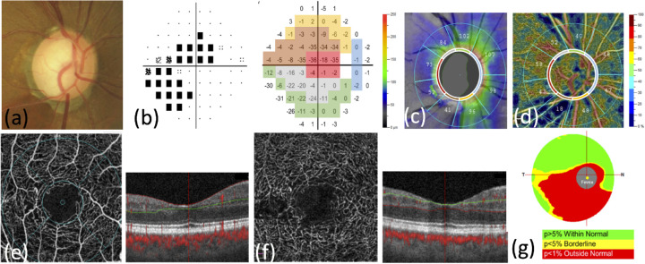
A total of 74 NTG eyes and 24 healthy eyes were included. OCTA VD at 4.5 × 4.5 mm peripapillary region and 3.0 × 3.0 mm macula area, RNFL thickness, and VF pattern deviation results were individually analyzed on the basis of the Garway-Heath sectorization. Correlations between VD and VF/RNFL and VF were compared.

RESULTS

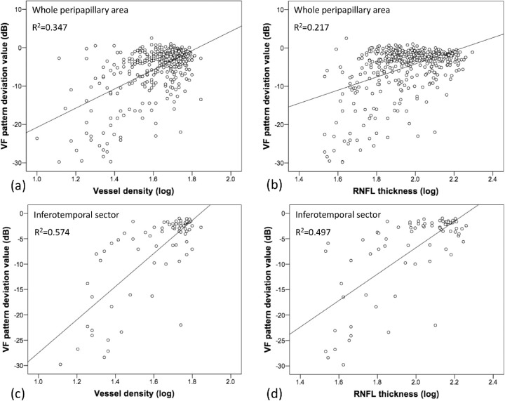
In the NTG group, peripapillary VD, superficial macula VD, RNFL thickness, and ganglion cell complex thickness were significantly lower. In the whole peripapillary area and inferotemporal sector, anatomic correlations between VD and VF pattern deviation values were significantly higher than those between the RNFL thickness and VF values. In the subgroup analysis, VD was anatomically correlated with VF in early-, moderate-, and severe-stage NTG eyes, whereas the RNFL thickness was correlated with VF in moderate- and severe-stage NTG eyes.

CONCLUSIONS

We observed VD reduction in the peripapillary retina and superficial macula area in NTG eyes. The microvascular dropout of VD in the peripapillary retina, especially in the inferotemporal sector, provided a more accurate anatomic correlation with functional VF defects than that of the RNFL thickness, especially in early-stage NTG eyes.

TRANSLATIONAL RELEVANCE

In patients who cannot comply VF exam, VD is a good tool for disease detection.

摘要

目的

通过光学相干断层扫描血管造影(OCTA)研究健康眼和正常眼压性青光眼(NTG)眼中的视网膜血管密度(VD),并确定不同位置的VD与视网膜神经纤维层(RNFL)厚度及功能性视野(VF)缺损之间的相关性。

方法

共纳入74只NTG眼和24只健康眼。基于Garway-Heath扇形分区,分别分析视乳头周围4.5×4.5mm区域和黄斑区3.0×3.0mm的OCTA血管密度、RNFL厚度及VF模式偏差结果。比较VD与VF/RNFL以及VF之间的相关性。

结果

在NTG组中,视乳头周围VD、黄斑浅层VD、RNFL厚度和神经节细胞复合体厚度显著降低。在整个视乳头周围区域和颞下扇形区,VD与VF模式偏差值之间的解剖学相关性显著高于RNFL厚度与VF值之间的相关性。在亚组分析中,早期、中期和重度NTG眼中VD与VF存在解剖学相关性,而中期和重度NTG眼中RNFL厚度与VF相关。

结论

我们观察到NTG眼中视乳头周围视网膜和黄斑浅层区域的VD降低。视乳头周围视网膜中VD的微血管缺失,尤其是在颞下扇形区,与功能性VF缺损的解剖学相关性比RNFL厚度更准确,尤其是在早期NTG眼中。

转化相关性

对于无法配合VF检查的患者,VD是疾病检测的良好工具。

https://cdn.ncbi.nlm.nih.gov/pmc/blobs/9c24/7746955/2461d638780c/tvst-9-13-26-f002.jpg
https://cdn.ncbi.nlm.nih.gov/pmc/blobs/9c24/7746955/486812c4bd98/tvst-9-13-26-f001.jpg
https://cdn.ncbi.nlm.nih.gov/pmc/blobs/9c24/7746955/2461d638780c/tvst-9-13-26-f002.jpg
https://cdn.ncbi.nlm.nih.gov/pmc/blobs/9c24/7746955/486812c4bd98/tvst-9-13-26-f001.jpg
https://cdn.ncbi.nlm.nih.gov/pmc/blobs/9c24/7746955/2461d638780c/tvst-9-13-26-f002.jpg

相似文献

1
Correlation of Visual Field With Peripapillary Vessel Density Through Optical Coherence Tomography Angiography in Normal-Tension Glaucoma.正常眼压性青光眼中通过光学相干断层扫描血管造影术评估视野与视乳头周围血管密度的相关性
Transl Vis Sci Technol. 2020 Dec 17;9(13):26. doi: 10.1167/tvst.9.13.26. eCollection 2020 Dec.
2
Longitudinal changes in optical coherence tomography angiography characteristics in normal-tension glaucoma with or without high myopia.正常眼压性青光眼伴或不伴高度近视的光学相干断层扫描血管造影特征的纵向变化。
Acta Ophthalmol. 2024 Aug;102(5):e762-e773. doi: 10.1111/aos.16644. Epub 2024 Jan 26.
3
[The function-structure impairment pattern of optic nerves in primary open-angle glaucoma and normal-tension glaucoma].[原发性开角型青光眼和正常眼压性青光眼视神经的功能-结构损害模式]
Zhonghua Yan Ke Za Zhi. 2018 Nov 11;54(11):811-819. doi: 10.3760/cma.j.issn.0412-4081.2018.11.004.
4
Optical Coherence Tomography Angiography Vessel Density Changes in Normal-tension Glaucoma Treated With Carteolol, Brimonidine, or Dorzolamide.正常眼压性青光眼经卡替洛尔、溴莫尼定或多佐胺治疗后的光学相干断层扫描血管密度变化。
J Glaucoma. 2021 Aug 1;30(8):690-696. doi: 10.1097/IJG.0000000000001859.
5
Optical coherence tomography angiography and the visual field in hypertensive and normotensive glaucoma.光学相干断层扫描血管造影与高血压和正常眼压性青光眼的视野。
Biomed Pap Med Fac Univ Palacky Olomouc Czech Repub. 2021 Nov;165(4):441-444. doi: 10.5507/bp.2020.044. Epub 2020 Oct 15.
6
Optical Coherence Tomography Angiography of Optic Disc in Eyes With Primary Open-angle Glaucoma and Normal-tension Glaucoma.原发性开角型青光眼和正常眼压性青光眼患者视盘的光学相干断层扫描血管造影。
J Glaucoma. 2019 Mar;28(3):243-251. doi: 10.1097/IJG.0000000000001184.
7
Comparison of Sectoral Structure-Function Relationships in Glaucoma: Vessel Density Versus Thickness in the Peripapillary Retinal Nerve Fiber Layer.青光眼各区域结构-功能关系的比较:视乳头周围视网膜神经纤维层的血管密度与厚度
Invest Ophthalmol Vis Sci. 2017 Oct 1;58(12):5251-5262. doi: 10.1167/iovs.17-21955.
8
Quantitative Analysis of Microvasculature in Macular and Peripapillary Regions in Early Primary Open-Angle Glaucoma.早期原发性开角型青光眼黄斑及视盘周围区域微血管定量分析。
Curr Eye Res. 2020 May;45(5):629-635. doi: 10.1080/02713683.2019.1676912. Epub 2019 Oct 14.
9
A Sectoral Analysis of Vessel Density Measurements in Perimetrically Intact Regions of Glaucomatous Eyes: An Optical Coherence Tomography Angiography Study.青光眼周边正常视野区血管密度的节段性分析:一项光学相干断层扫描血管造影研究。
J Glaucoma. 2018 Jun;27(6):525-531. doi: 10.1097/IJG.0000000000000950.
10
Peripapillary and Macular Vessel Density Measurement by Optical Coherence Tomography Angiography in Pseudoexfoliation and Primary Open-angle Glaucoma.光学相干断层扫描血管造影术在假性剥脱综合征和原发性开角型青光眼的周边和黄斑血管密度测量。
J Glaucoma. 2020 May;29(5):381-385. doi: 10.1097/IJG.0000000000001464.

引用本文的文献

1
Association of contrast sensitivity with vessel density and functional parameters in different stages of glaucoma.青光眼不同阶段对比敏感度与血管密度及功能参数的相关性
Graefes Arch Clin Exp Ophthalmol. 2025 Feb;263(2):477-487. doi: 10.1007/s00417-024-06626-3. Epub 2024 Sep 5.
2
Detection and agreement of event-based OCT and OCTA analysis for glaucoma progression.基于事件的 OCT 和 OCTA 分析检测和一致性在青光眼进展中的应用。
Eye (Lond). 2024 Apr;38(5):973-979. doi: 10.1038/s41433-023-02817-0. Epub 2023 Nov 11.
3
Optical Coherence Tomography Angiography (OCTA) Differences in Vessel Perfusion Density and Flux Index of the Optic Nerve and Peri-Papillary Area in Healthy, Glaucoma Suspect and Glaucomatous Eyes.

本文引用的文献

1
Parafoveal vessel changes in primary open-angle glaucoma and normal-tension glaucoma using optical coherence tomography angiography.使用光学相干断层扫描血管造影术观察原发性开角型青光眼和正常眼压性青光眼的黄斑旁血管变化
Clin Ophthalmol. 2019 Sep 27;13:1935-1945. doi: 10.2147/OPTH.S206288. eCollection 2019.
2
Early Macular Angiography among Patients with Glaucoma, Ocular Hypertension, and Normal Subjects.青光眼、高眼压症患者及正常受试者的早期黄斑血管造影
J Ophthalmol. 2019 Jan 15;2019:7419470. doi: 10.1155/2019/7419470. eCollection 2019.
3
Optical Coherence Tomography Angiography of Optic Disc in Eyes With Primary Open-angle Glaucoma and Normal-tension Glaucoma.
光学相干断层扫描血管造影(OCTA)在健康眼、青光眼可疑眼和青光眼眼中视神经及视乳头周围区域血管灌注密度和血流指数的差异。
Clin Ophthalmol. 2023 Oct 12;17:3011-3021. doi: 10.2147/OPTH.S429718. eCollection 2023.
4
Peripapillary Vascular Density in Compressive Optic Neuropathy and Normal-Tension Glaucoma: A Severity-Controlled Comparison.压迫性视神经病变和正常眼压性青光眼的视盘旁血管密度:严重程度对照比较。
Invest Ophthalmol Vis Sci. 2023 Sep 1;64(12):10. doi: 10.1167/iovs.64.12.10.
5
Neuroprotection in Glaucoma: Basic Aspects and Clinical Relevance.青光眼的神经保护:基础方面与临床相关性
J Pers Med. 2022 Nov 10;12(11):1884. doi: 10.3390/jpm12111884.
6
The Topographic Relationship Between Choroidal Microvascular Dropout and Glaucomatous Damage in Primary Angle-Closure Glaucoma.原发性闭角型青光眼脉络膜小血管丢失与青光眼损害的位置关系。
Transl Vis Sci Technol. 2022 Oct 3;11(10):20. doi: 10.1167/tvst.11.10.20.
7
A Global and Sector-Based Comparison of OCT Angiography and Visual Field Defects in Glaucoma.青光眼患者光学相干断层扫描血管造影与视野缺损的全球及基于疾病领域的比较
J Ophthalmol. 2022 May 11;2022:6182592. doi: 10.1155/2022/6182592. eCollection 2022.
原发性开角型青光眼和正常眼压性青光眼患者视盘的光学相干断层扫描血管造影。
J Glaucoma. 2019 Mar;28(3):243-251. doi: 10.1097/IJG.0000000000001184.
4
Diagnostic Performance of Macular Versus Peripapillary Vessel Parameters by Optical Coherence Tomography Angiography for Glaucoma.光学相干断层扫描血管造影术检测黄斑与视乳头周围血管参数对青光眼的诊断效能
Transl Vis Sci Technol. 2018 Dec 6;7(6):21. doi: 10.1167/tvst.7.6.21. eCollection 2018 Nov.
5
Optical Coherence Tomography Angiography in Glaucoma: A Review.青光眼的光学相干断层扫描血管造影:综述
Ophthalmic Res. 2018;60(3):139-151. doi: 10.1159/000488495. Epub 2018 May 24.
6
Optical Coherence Tomography Angiography Macular Vascular Density Measurements and the Central 10-2 Visual Field in Glaucoma.光学相干断层扫描血管造影黄斑血管密度测量与青光眼中心 10-2 视野。
J Glaucoma. 2018 Jun;27(6):481-489. doi: 10.1097/IJG.0000000000000964.
7
Comparison of Optical Coherence Tomography Angiography and Laser Speckle Flowgraphy for the Diagnosis of Normal-Tension Glaucoma.光学相干断层扫描血管造影术与激光散斑血流图在正常眼压性青光眼诊断中的比较
J Ophthalmol. 2018 Jan 31;2018:1751857. doi: 10.1155/2018/1751857. eCollection 2018.
8
The Association Between Macula and ONH Optical Coherence Tomography Angiography (OCT-A) Vessel Densities in Glaucoma, Glaucoma Suspect, and Healthy Eyes.青光眼、疑似青光眼和健康眼中黄斑和视盘 OCT-A 血管密度的相关性。
J Glaucoma. 2018 Mar;27(3):227-232. doi: 10.1097/IJG.0000000000000862.
9
Progressive Macula Vessel Density Loss in Primary Open-Angle Glaucoma: A Longitudinal Study.原发性开角型青光眼黄斑区血管密度的渐进性丧失:一项纵向研究。
Am J Ophthalmol. 2017 Oct;182:107-117. doi: 10.1016/j.ajo.2017.07.011. Epub 2017 Jul 20.
10
[Optical coherence tomography angiography in glaucoma diagnosis].[光学相干断层扫描血管造影在青光眼诊断中的应用]
Vestn Oftalmol. 2016;132(5):98-102. doi: 10.17116/oftalma2016132598-102.