Yonemizu Sayaka, Masuda Keiichiro, Kurata Yasutaka, Notsu Tomomi, Higashi Yuhei, Fukumura Kenta, Li Peili, Ninomiya Haruaki, Miake Junichiro, Tsuneto Motokazu, Shirayoshi Yasuaki, Hisatome Ichiro
Division of Regenerative Medicine and Therapeutics, Department of Genetic Medicine and Regenerative Therapeutics, Tottori University Graduate School of Medical Science, 86 Nishi-cho, Yonago 683-8503, Japan.
Department of Physiology II, Kanazawa Medical University, 1-1 Daigaku, Uchinada-machi, Kahoku-gun, Ishikawa 920-0293, Japan.
Regen Ther. 2019 Feb 1;10:104-111. doi: 10.1016/j.reth.2018.12.002. eCollection 2019 Jun.
Human induced pluripotent stem cells (hiPSCs) harboring cardiac myosin heavy chain 6 promoter can differentiate into functional cardiomyocytes called "iCell cardiomyocytes" under blasticidin treatment condition. While iCell cardiomyocytes are expected to be used for predicting cardiotoxicity of drugs, their responses to antiarrhythmic agents remain to be elucidated. We first examined electrophysiological properties of iCell cardiomyocytes and mRNA levels of ion channels and Ca handling proteins, and then evaluated effects of class I antiarrhythmic agents on their Na and Ca currents.
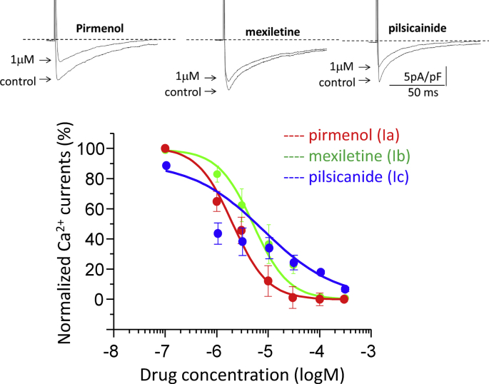
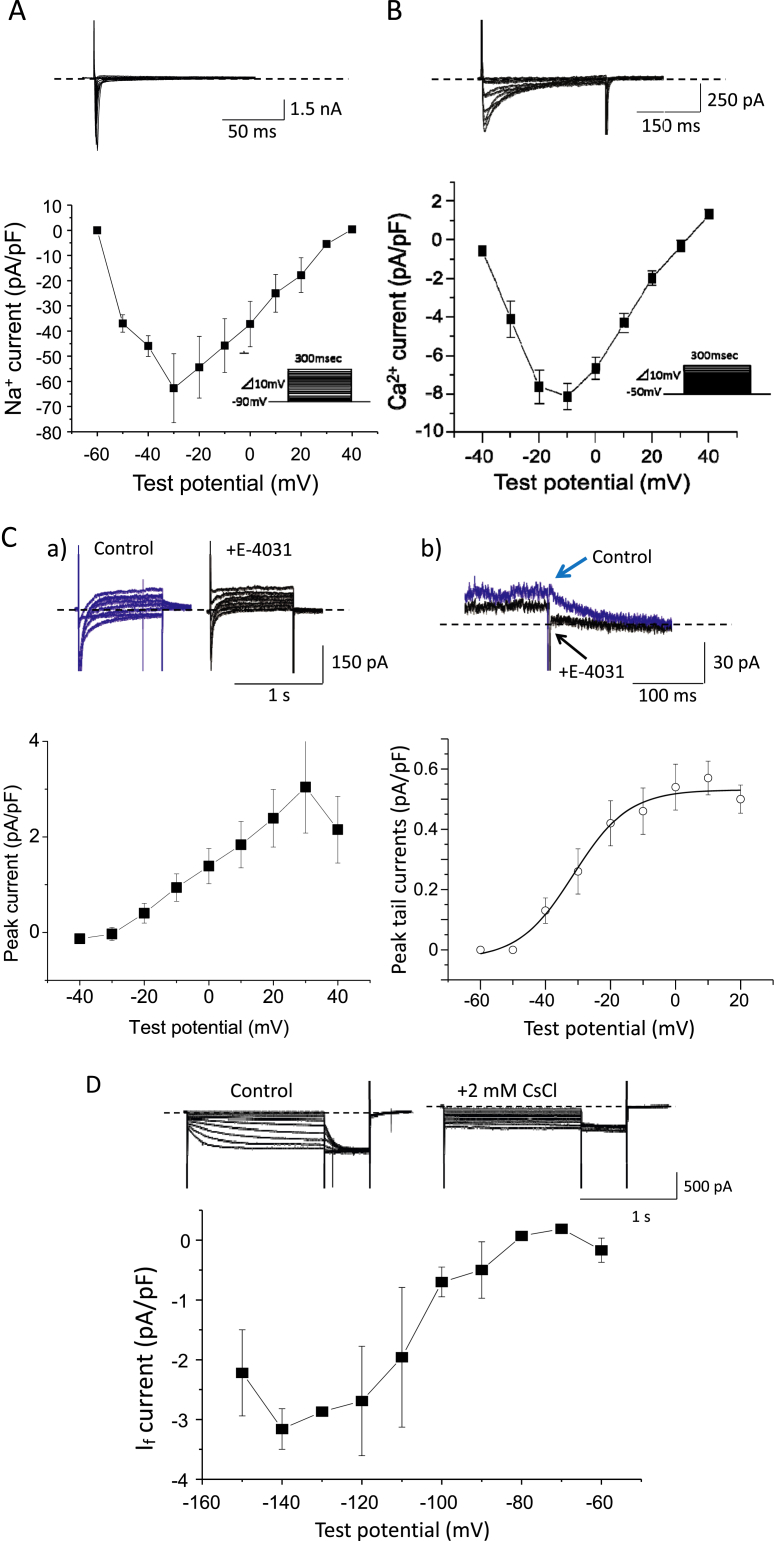
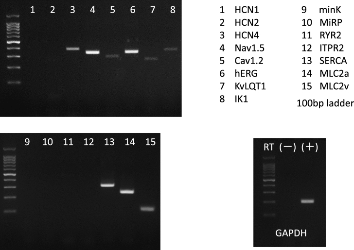
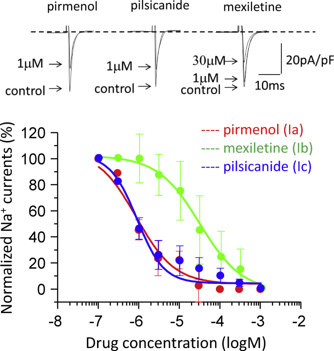
iCell cardiomyocytes were cultured for 8-14 days (38-44 days after inducing their differentiation), according to the manufacturer's protocol. We determined their action potentials (APs) and sarcolemmal ionic currents using whole-cell patch clamp techniques, and also mRNA levels of ion channels and Ca handling proteins by RT-PCR. Effects of three class I antiarrhythmic agents, pirmenol, pilsicainide and mexiletine, on Na channel current (I) and L-type Ca channel current (I) were evaluated by the whole-cell patch clamp.
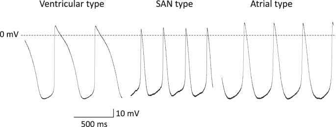
iCell cardiomyocytes revealed sinoatrial node-type (18%), atrial-type (18%) and ventricular-type (64%) spontaneous APs. The maximum peak amplitudes of I, I, and rapidly-activating delayed-rectifier K channel current were -62.7 ± 13.7, -8.1 ± 0.7, and 3.0 ± 1.0 pA/pF, respectively. The hyperpolarization-activated cation channel and inward-rectifier K channel currents were observed, whereas the T-type Ca channel or slowly-activating delayed-rectifier K channel current was not detectable. mRNAs of Nav1.5, Cav1.2, Kir2.1, HCN4, KvLQT1, hERG and SERCA2 were detected, while that of HCN1, minK or MiRP was not. The class Ia antiarrhythmic agent pirmenol and class Ic agent pilsicainide blocked I in a concentration-dependent manner with IC of 0.87 ± 0.37 and 0.88 ± 0.16 μM, respectively; the class Ib agent mexiletine revealed weak I block with a higher IC of 30.0 ± 3.0 μM. Pirmenol, pilsicainide and mexiletine blocked I with IC of 2.00 ± 0.39, 7.7 ± 2.5 and 5.0 ± 0.1 μM, respectively.
In iCell cardiomyocytes, I was blocked by the class Ia and Ic antiarrhythmic agents and I was blocked by all the class I agents within the ranges of clinical concentrations, suggesting their cardiotoxicity.
携带心肌肌球蛋白重链6启动子的人诱导多能干细胞(hiPSC)在杀稻瘟菌素处理条件下可分化为功能性心肌细胞,称为“iCell心肌细胞”。虽然iCell心肌细胞有望用于预测药物的心脏毒性,但其对抗心律失常药物的反应仍有待阐明。我们首先检测了iCell心肌细胞的电生理特性以及离子通道和钙处理蛋白的mRNA水平,然后评估了I类抗心律失常药物对其钠电流和钙电流的影响。
根据制造商的方案,将iCell心肌细胞培养8 - 14天(诱导分化后38 - 44天)。我们使用全细胞膜片钳技术测定其动作电位(AP)和肌膜离子电流,并通过逆转录聚合酶链反应(RT-PCR)检测离子通道和钙处理蛋白的mRNA水平。通过全细胞膜片钳评估三种I类抗心律失常药物,吡美诺、吡西卡尼和美西律对钠通道电流(I)和L型钙通道电流(I)的影响。
iCell心肌细胞表现出窦房结型(18%)、心房型(18%)和心室型(64%)的自发动作电位。I、I和快速激活延迟整流钾通道电流的最大峰值幅度分别为-62.7±13.7、-8.1±0.7和3.0±1.0 pA/pF。观察到超极化激活阳离子通道和内向整流钾通道电流,而未检测到T型钙通道或缓慢激活延迟整流钾通道电流。检测到Nav1.5、Cav1.2、Kir2.1、HCN4、KvLQT1、hERG和SERCA2的mRNA,而未检测到HCN1、minK或MiRP的mRNA。I类a抗心律失常药物吡美诺和I类c药物吡西卡尼以浓度依赖性方式阻断I,IC分别为0.87±0.37和0.88±0.16μM;I类b药物美西律显示出较弱地阻断I,IC较高,为30.0±3.0μM。吡美诺、吡西卡尼和美西律阻断I的IC分别为2.00±0.39、7.7±2.5和5.0±0.1μM。
在iCell心肌细胞中,I类a和I类c抗心律失常药物在临床浓度范围内阻断I,所有I类药物在临床浓度范围内阻断I,提示它们具有心脏毒性。