The Mood and Anxiety Disorders Program, Department of Psychiatry, The Icahn School of Medicine at Mount Sinai, New York, NY, 10029, USA.
The Translational and Molecular Imaging Institute, Department of Radiology, The Icahn School of Medicine at Mount Sinai, New York, NY, 10029, USA.
Transl Psychiatry. 2019 Feb 15;9(1):94. doi: 10.1038/s41398-019-0425-6.
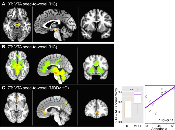
Ultra-high field 7-Tesla (7 T) MRI has the potential to advance our understanding of neuropsychiatric disorders, including major depressive disorder (MDD). To date, few studies have quantified the advantage of resting state functional MRI (fMRI) at 7 T compared to 3-Tesla (3 T). We conducted a series of experiments that demonstrate the improvement in temporal signal-to-noise ratio (TSNR) of a multi-echo multi-band fMRI protocol with ultra-high field 7 T MRI, compared to a similar protocol using 3 T MRI in healthy controls (HC). We also directly tested the enhancement in ultra-high field 7 T fMRI signal power by examining the ventral tegmental area (VTA), a small midbrain structure that is critical to the expected neuropathology of MDD but difficult to discern with standard 3 T MRI. We demonstrate up to 300% improvement in TSNR and resting state functional connectivity coefficients provided by ultra-high field 7 T fMRI compared to 3 T, indicating enhanced power for detection of functional neural architecture. A multi-echo based acquisition protocol and signal denoising pipeline afforded greater gain in signal power compared to classic acquisition and denoising pipelines. Furthermore, ultra-high field fMRI revealed mood-related neurocircuit disturbances in patients with MDD compared to HC, which were not detectable with 3 T fMRI. Ultra-high field 7 T fMRI may provide an effective tool for studying functional neural architecture relevant to MDD and other neuropsychiatric disorders.
超高场 7 特斯拉(7T)磁共振成像有可能增进我们对神经精神疾病的理解,包括重度抑郁症(MDD)。迄今为止,很少有研究定量比较了 7T 静息态功能磁共振成像(fMRI)与 3T 的优势。我们进行了一系列实验,证明了多回波多频带 fMRI 方案在超高场 7T 磁共振成像中的时间信号噪声比(TSNR)改善,与在健康对照组(HC)中使用 3T MRI 的类似方案相比。我们还通过检查腹侧被盖区(VTA)直接测试了超高场 7T fMRI 信号功率的增强,VTA 是一个小脑结构,对 MDD 的预期神经病理学至关重要,但用标准的 3T MRI 难以辨别。与 3T 相比,我们证明了超高场 7T fMRI 的 TSNR 和静息态功能连接系数提高了高达 300%,表明检测功能神经结构的功率增强。与经典采集和降噪方案相比,基于多回波的采集方案和信号降噪处理管道提供了更大的信号功率增益。此外,与 3T fMRI 相比,超高场 fMRI 揭示了 MDD 患者与 HC 相比存在与情绪相关的神经回路紊乱,而这些紊乱是无法用 3T fMRI 检测到的。超高场 7T fMRI 可能成为研究与 MDD 和其他神经精神疾病相关的功能神经结构的有效工具。