Li Xian, Chen Shurui, Mao Liang, Li Daoyong, Xu Chang, Tian He, Mei Xifan
Department of Orthopedics, The First Affiliated Hospital of Jinzhou Medical University, Jinzhou, China.
Department of Oncology, The First Affiliated Hospital of Jinzhou Medical University, Jinzhou, China.
Front Mol Neurosci. 2019 Feb 1;12:18. doi: 10.3389/fnmol.2019.00018. eCollection 2019.
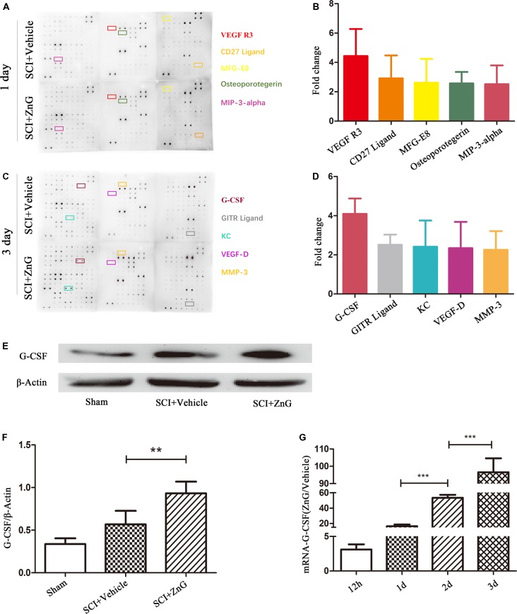
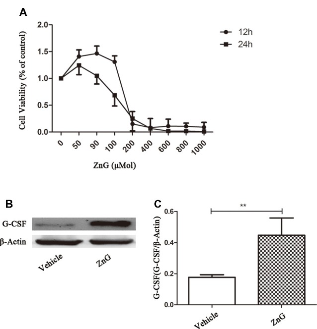
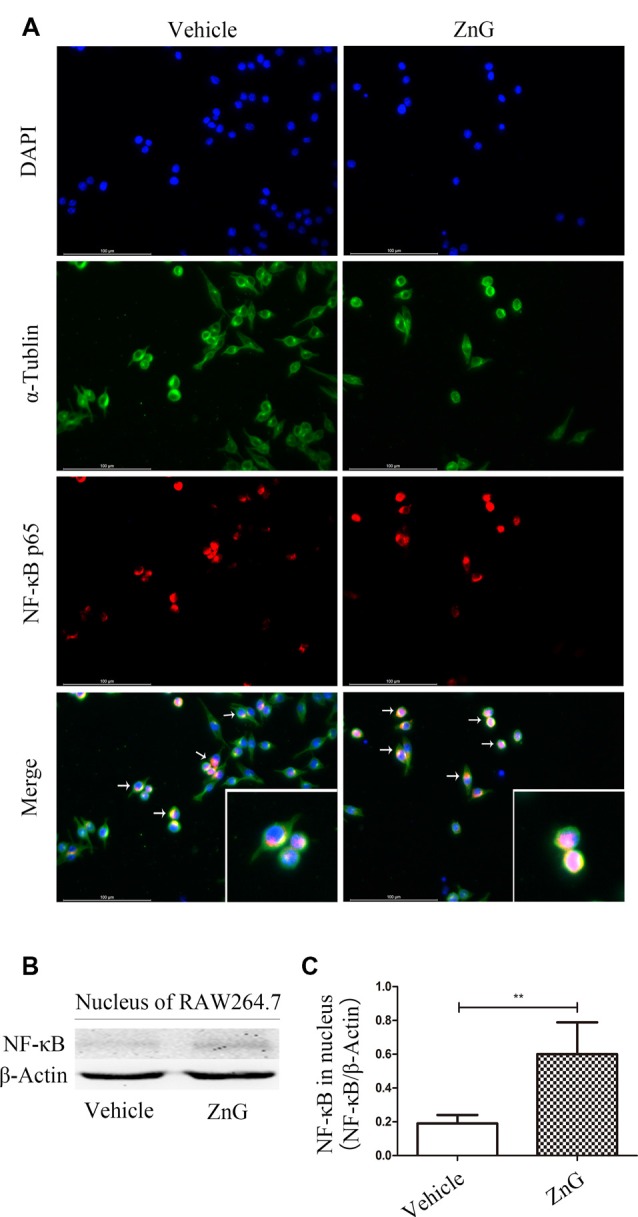
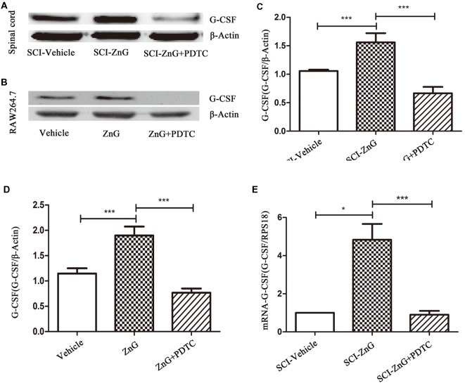
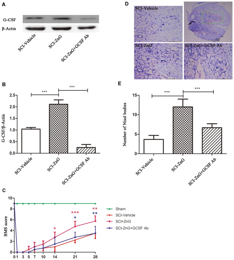
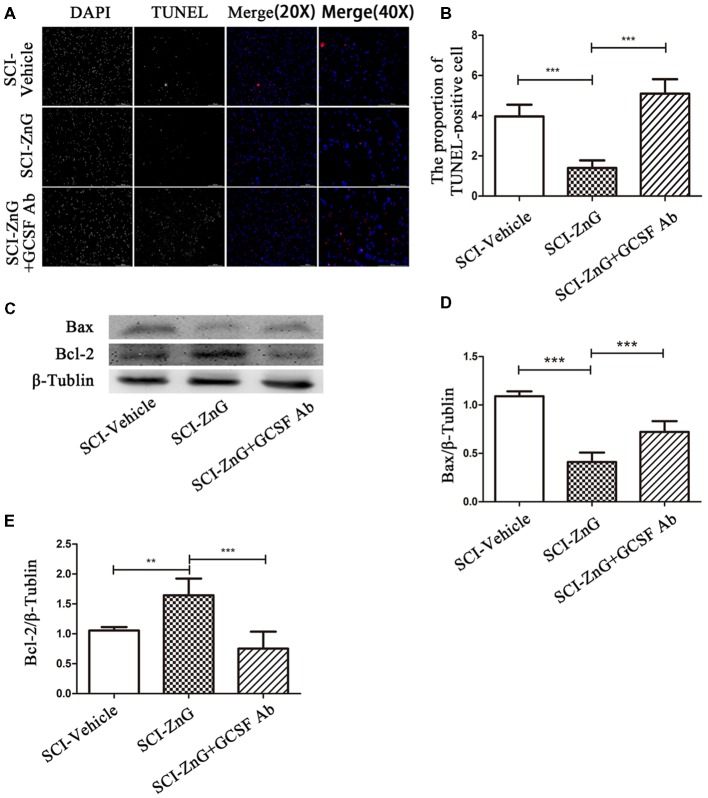
While zinc promotes motor function recovery after spinal cord injury (SCI), the precise mechanisms involved are not fully understood. The present study aimed to elucidate the effects of zinc and granulocyte colony stimulating factor (G-CSF) on neuronal recovery after SCI. The SCI model was established by Allen's method. Injured animals were given glucose and zinc gluconate (ZnG; 30 mg/kg) for the first time at 2 h after injury, the same dose was given for 3 days. A cytokine antibody array was used to screen changes in inflammation at the site of SCI lesion. Immunofluorescence was used to detect the distribution of cytokines. Magnetic beads were also used to isolate cells from the site of SCI lesion. We then investigated the effect of Zinc on apoptosis after SCI by Transferase UTP Nick End Labeling (TUNEL) staining and Western Blotting. Basso Mouse Scale (BMS) scores and immunofluorescence were employed to investigate neuronal apoptosis and functional recovery. We found that the administration of zinc significantly increased the expression of 19 cytokines in the SCI lesion. Of these, G-CSF was shown to be the most elevated cytokine and was secreted by microglia/macrophages (M/Ms) the nuclear factor-kappa B (NF-κB) signaling pathway after SCI. Increased levels of G-CSF at the SCI lesion reduced the level of neuronal apoptosis after SCI, thus promoting functional recovery. Collectively, our results indicate that the administration of zinc increases the expression of G-CSF secreted by M/Ms, which then leads to reduced levels of neuronal apoptosis after SCI.
虽然锌可促进脊髓损伤(SCI)后运动功能的恢复,但其具体机制尚未完全明确。本研究旨在阐明锌和粒细胞集落刺激因子(G-CSF)对SCI后神经元恢复的影响。采用Allen法建立SCI模型。受伤动物在损伤后2小时首次给予葡萄糖和葡萄糖酸锌(ZnG;30mg/kg),连续3天给予相同剂量。使用细胞因子抗体阵列筛选SCI损伤部位炎症的变化。采用免疫荧光法检测细胞因子的分布。还使用磁珠从SCI损伤部位分离细胞。然后,我们通过末端脱氧核苷酸转移酶介导的缺口末端标记(TUNEL)染色和蛋白质免疫印迹法研究锌对SCI后细胞凋亡的影响。采用Basso小鼠评分(BMS)和免疫荧光法研究神经元凋亡和功能恢复情况。我们发现,给予锌可显著增加SCI损伤部位19种细胞因子的表达。其中,G-CSF是表达上调最为明显的细胞因子,由小胶质细胞/巨噬细胞(M/Ms)在SCI后通过核因子-κB(NF-κB)信号通路分泌。SCI损伤部位G-CSF水平的升高降低了SCI后神经元凋亡水平,从而促进功能恢复。总的来说,我们的结果表明,给予锌可增加M/Ms分泌的G-CSF的表达,进而降低SCI后神经元凋亡水平。