• 文献检索
  • 文档翻译
  • 深度研究
  • 学术资讯
  • Suppr Zotero 插件Zotero 插件
  • 邀请有礼
  • 套餐&价格
  • 历史记录
应用&插件
Suppr Zotero 插件Zotero 插件浏览器插件Mac 客户端Windows 客户端微信小程序
定价
高级版会员购买积分包购买API积分包
服务
文献检索文档翻译深度研究API 文档MCP 服务
关于我们
关于 Suppr公司介绍联系我们用户协议隐私条款
关注我们

Suppr 超能文献

核心技术专利:CN118964589B侵权必究
粤ICP备2023148730 号-1Suppr @ 2026

文献检索

告别复杂PubMed语法,用中文像聊天一样搜索,搜遍4000万医学文献。AI智能推荐,让科研检索更轻松。

立即免费搜索

文件翻译

保留排版,准确专业,支持PDF/Word/PPT等文件格式,支持 12+语言互译。

免费翻译文档

深度研究

AI帮你快速写综述,25分钟生成高质量综述,智能提取关键信息,辅助科研写作。

立即免费体验

基于激光的 3D 生物打印用于肿瘤球体和类胚胎体的空间和大小控制。

Laser-based 3D bioprinting for spatial and size control of tumor spheroids and embryoid bodies.

机构信息

Department of Biomedical Engineering, Rensselaer Polytechnic Institute, 110 Eighth St., Troy, NY 12180, USA.

Department of Molecular and Cellular Physiology, Albany Medical College, Albany, NY 12208, USA.

出版信息

Acta Biomater. 2019 Sep 1;95:357-370. doi: 10.1016/j.actbio.2019.02.014. Epub 2019 Feb 15.

DOI:10.1016/j.actbio.2019.02.014
PMID:30776506
原文链接:https://pmc.ncbi.nlm.nih.gov/articles/PMC7171976/
Abstract

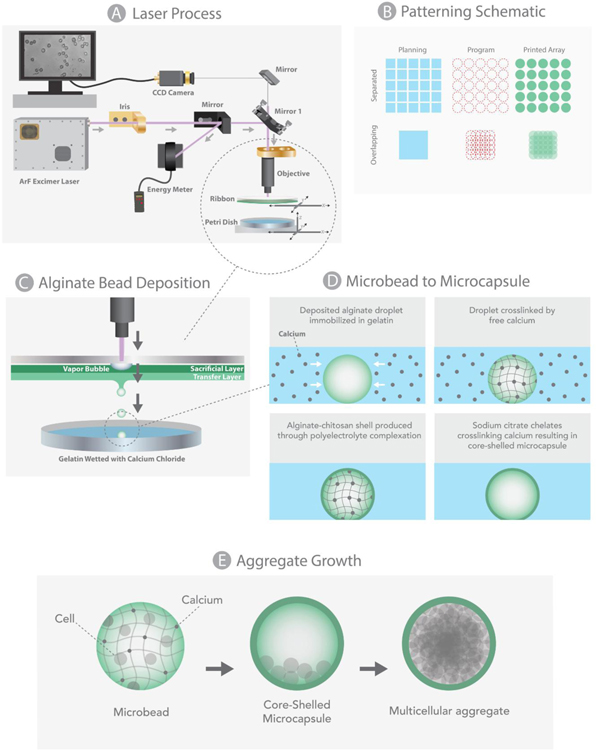
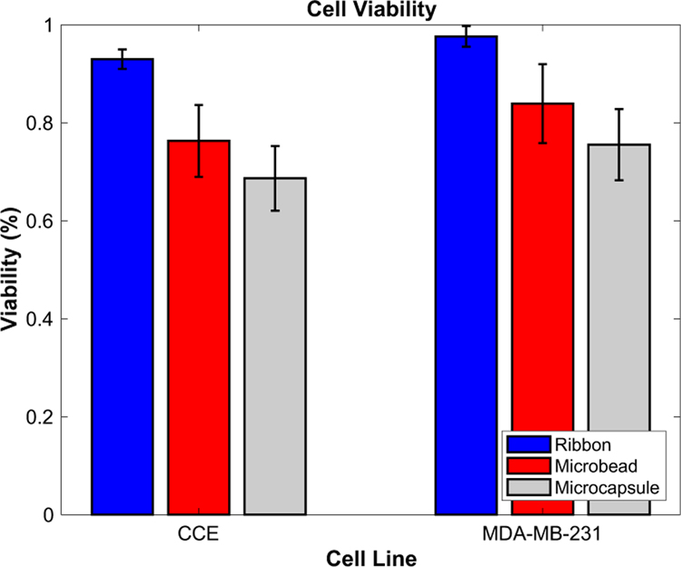
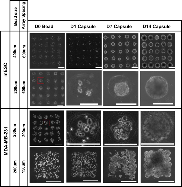
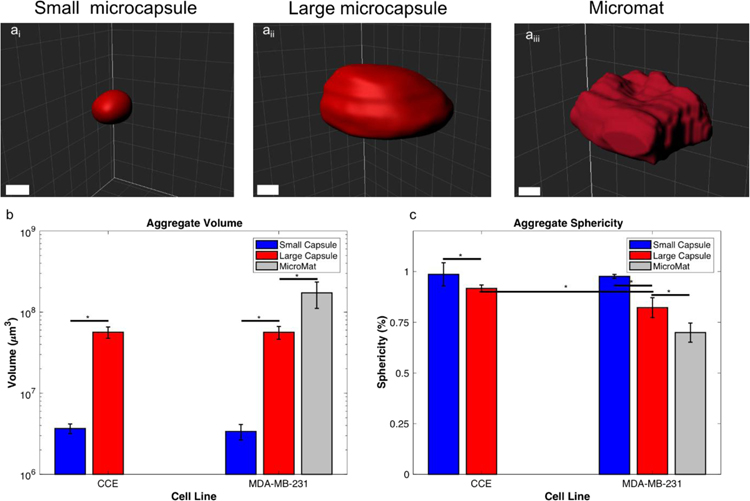
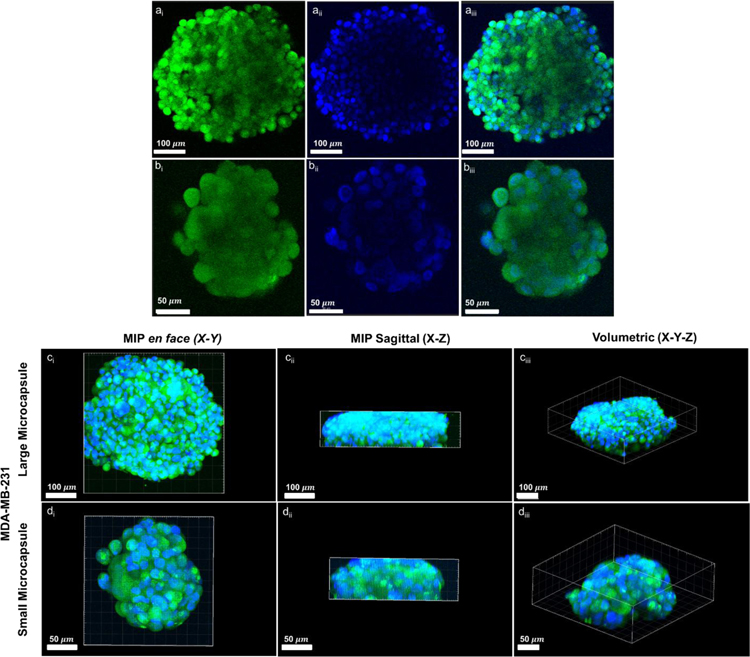
3D multicellular aggregates, and more advanced organotypic systems, have become central tools in recent years to study a wide variety of complex biological processes. Most notably, these model systems have become mainstream within oncology (multicellular tumor spheroids) and regenerative medicine (embryoid bodies) research. However, the biological behavior of these in vitro tissue surrogates is extremely sensitive to their aggregate size and geometry. Indeed, both of these geometrical parameters are key in producing pathophysiological gradients responsible for cellular and structural heterogeneity, replicating in vivo observations. Moreover, the fabrication techniques most widely used for producing these models lack the ability to accurately control cellular spatial location, an essential component for regulating homotypic and heterotypic cell signaling. Herein, we report on a 3D bioprinting technique, laser direct-write (LDW), that enables precise control of both spatial patterning and size of cell-encapsulating microbeads. The generated cell-laden beads are further processed into core-shelled structures, allowing for the growth and formation of self-contained, self-aggregating cells (e.g., breast cancer cells, embryonic stem cells). Within these structures we demonstrate our ability to produce multicellular tumor spheroids (MCTSs) and embryoid bodies (EBs) with well-controlled overall size and shape, that can be designed on demand. Furthermore, we investigated the impact of aggregate size on the uptake of a commonly employed ligand for receptor-mediated drug delivery, Transferrin, indicating that larger tumor spheroids exhibit greater spatial heterogeneity in ligand uptake. Taken together, these findings establish LDW as a versatile biomanufacturing platform for bioprinting and patterning core-shelled structures to generate size-controlled 3D multicellular aggregates. STATEMENT OF SIGNIFICANCE: Multicellular 3D aggregates are powerful in vitro models used to study a wide variety of complex biological processes, particularly within oncology and regenerative medicine. These tissue surrogates are fabricated using environments that encourage cellular self-assembly. However, specific applications require control of aggregate size and position to recapitulate key in vivo parameters (e.g., pathophysiological gradients and homotypic/heterotypic cell signaling). Herein, we demonstrate the ability to create and spatially pattern size-controlled embryoid bodies and tumor spheroids, using laser-based 3D bioprinting. Furthermore, we investigated the effect of tumor spheroid size on internalization of Transferrin, a common ligand for targeted therapy, finding greater spatial heterogeneity in our large aggregates. Overall, this technique offers incredible promise and flexibility for fabricating idealized 3D in vitro models.

摘要

3D 多细胞聚集体和更先进的器官型系统近年来已成为研究各种复杂生物过程的重要工具。特别是,这些模型系统已成为肿瘤学(多细胞肿瘤球体)和再生医学(胚胎体)研究的主流。然而,这些体外组织替代物的生物学行为对其聚集体的大小和几何形状极为敏感。事实上,这两个几何参数对于产生负责细胞和结构异质性的病理生理梯度至关重要,从而复制体内观察结果。此外,用于生产这些模型的最广泛使用的制造技术缺乏准确控制细胞空间位置的能力,而细胞空间位置是调节同型和异型细胞信号传导的重要组成部分。在这里,我们报告了一种 3D 生物打印技术,即激光直写(LDW),该技术可以精确控制细胞包封微珠的空间图案和大小。生成的细胞负载珠进一步加工成核壳结构,允许自包含、自聚集细胞(例如乳腺癌细胞、胚胎干细胞)的生长和形成。在这些结构中,我们展示了我们生产具有良好控制的整体大小和形状的多细胞肿瘤球体(MCTS)和胚胎体(EBs)的能力,可以根据需要进行设计。此外,我们研究了聚集体大小对常用于受体介导药物输送的配体转铁蛋白摄取的影响,结果表明较大的肿瘤球体在配体摄取方面表现出更大的空间异质性。总之,这些发现确立了 LDW 作为一种用于生物打印和图案化核壳结构以生成尺寸可控的 3D 多细胞聚集体的多功能生物制造平台。

意义声明

多细胞 3D 聚集体是用于研究各种复杂生物过程的强大体外模型,特别是在肿瘤学和再生医学领域。这些组织替代物是使用促进细胞自组装的环境制造的。然而,特定应用需要控制聚集体的大小和位置,以再现关键的体内参数(例如,病理生理梯度和同型/异型细胞信号传导)。在这里,我们使用基于激光的 3D 生物打印演示了创建和空间图案化尺寸可控的胚胎体和肿瘤球体的能力。此外,我们研究了肿瘤球体大小对转铁蛋白摄取的影响,转铁蛋白是靶向治疗的常见配体,结果发现我们的大聚集体中存在更大的空间异质性。总体而言,该技术为制造理想的 3D 体外模型提供了令人难以置信的承诺和灵活性。

https://cdn.ncbi.nlm.nih.gov/pmc/blobs/8bb4/7171976/e0010e34e024/nihms-1524155-f0010.jpg
https://cdn.ncbi.nlm.nih.gov/pmc/blobs/8bb4/7171976/ab82854a69fc/nihms-1524155-f0002.jpg
https://cdn.ncbi.nlm.nih.gov/pmc/blobs/8bb4/7171976/3a63c10ba708/nihms-1524155-f0003.jpg
https://cdn.ncbi.nlm.nih.gov/pmc/blobs/8bb4/7171976/0f47ebd1b33c/nihms-1524155-f0004.jpg
https://cdn.ncbi.nlm.nih.gov/pmc/blobs/8bb4/7171976/50ad7c1d9fa8/nihms-1524155-f0005.jpg
https://cdn.ncbi.nlm.nih.gov/pmc/blobs/8bb4/7171976/32a1a0c56441/nihms-1524155-f0006.jpg
https://cdn.ncbi.nlm.nih.gov/pmc/blobs/8bb4/7171976/e668db5d5875/nihms-1524155-f0007.jpg
https://cdn.ncbi.nlm.nih.gov/pmc/blobs/8bb4/7171976/ea4135a5ec14/nihms-1524155-f0008.jpg
https://cdn.ncbi.nlm.nih.gov/pmc/blobs/8bb4/7171976/2f16739e109d/nihms-1524155-f0009.jpg
https://cdn.ncbi.nlm.nih.gov/pmc/blobs/8bb4/7171976/e0010e34e024/nihms-1524155-f0010.jpg

相似文献

1
Laser-based 3D bioprinting for spatial and size control of tumor spheroids and embryoid bodies.基于激光的 3D 生物打印用于肿瘤球体和类胚胎体的空间和大小控制。
Acta Biomater. 2019 Sep 1;95:357-370. doi: 10.1016/j.actbio.2019.02.014. Epub 2019 Feb 15.
2
Three-dimensional bioprinting of embryonic stem cells directs highly uniform embryoid body formation.胚胎干细胞的三维生物打印引导高度均匀的胚状体形成。
Biofabrication. 2015 Nov 4;7(4):044101. doi: 10.1088/1758-5090/7/4/044101.
3
Nonlinear 3D projection printing of concave hydrogel microstructures for long-term multicellular spheroid and embryoid body culture.用于长期细胞球状体和类胚胎体培养的凹形水凝胶微结构的非线性 3D 投影打印。
Lab Chip. 2015 Jun 7;15(11):2412-8. doi: 10.1039/c5lc00159e. Epub 2015 Apr 22.
4
Laser-assisted bioprinting of targeted cartilaginous spheroids for high density bottom-up tissue engineering.激光辅助靶向软骨球的生物打印在高密度自下而上的组织工程中的应用。
Biofabrication. 2024 Aug 22;16(4). doi: 10.1088/1758-5090/ad6e1a.
5
Generating size-controlled embryoid bodies using laser direct-write.利用激光直写技术生成尺寸可控的胚状体。
Biofabrication. 2014 Jun;6(2):025007. doi: 10.1088/1758-5082/6/2/025007. Epub 2014 Apr 3.
6
Direct Bioprinting of 3D Multicellular Breast Spheroids onto Endothelial Networks.将3D多细胞乳腺球体直接生物打印到内皮网络上。
J Vis Exp. 2020 Nov 2(165). doi: 10.3791/61791.
7
Strategies for 3D bioprinting of spheroids: A comprehensive review.球形聚集体的 3D 生物打印策略:全面综述。
Biomaterials. 2022 Dec;291:121881. doi: 10.1016/j.biomaterials.2022.121881. Epub 2022 Oct 28.
8
Aspiration-assisted bioprinting for precise positioning of biologics.辅助抽吸生物打印技术实现生物制剂的精准定位。
Sci Adv. 2020 Mar 6;6(10):eaaw5111. doi: 10.1126/sciadv.aaw5111. eCollection 2020 Mar.
9
In vitro spatially organizing the differentiation in individual multicellular stem cell aggregates.在体外空间组织个体多能干细胞聚集体的分化。
Crit Rev Biotechnol. 2016;36(1):20-31. doi: 10.3109/07388551.2014.922917. Epub 2014 Jul 15.
10
3D bioprinting of a stem cell-laden, multi-material tubular composite: An approach for spinal cord repair.负载干细胞的多材料管状复合材料的3D生物打印:一种脊髓修复方法。
Mater Sci Eng C Mater Biol Appl. 2021 Jan;120:111707. doi: 10.1016/j.msec.2020.111707. Epub 2020 Nov 6.

引用本文的文献

1
Exploring the Complexity of Cutaneous Squamous CellCarcinoma Microenvironment: Focus on Immune Cell Roles by Novel 3D In Vitro Models.探索皮肤鳞状细胞癌微环境的复杂性:通过新型三维体外模型聚焦免疫细胞的作用
Life (Basel). 2025 Jul 23;15(8):1170. doi: 10.3390/life15081170.
2
Tumor-infiltrating lymphocytes in cancer immunotherapy: from chemotactic recruitment to translational modeling.癌症免疫治疗中的肿瘤浸润淋巴细胞:从趋化募集到转化模型
Front Immunol. 2025 May 22;16:1601773. doi: 10.3389/fimmu.2025.1601773. eCollection 2025.
3
Modeling intratumor heterogeneity in breast cancer.

本文引用的文献

1
Advanced Cell and Tissue Biomanufacturing.先进细胞与组织生物制造
ACS Biomater Sci Eng. 2018 Jul 9;4(7):2292-2307. doi: 10.1021/acsbiomaterials.8b00650. Epub 2018 Jun 19.
2
An Ex Vivo Vessel Injury Model to Study Remodeling.用于研究重塑的离体血管损伤模型。
Cell Transplant. 2018 Sep;27(9):1375-1389. doi: 10.1177/0963689718792201. Epub 2018 Aug 10.
3
Laser bioprinting of human induced pluripotent stem cells-the effect of printing and biomaterials on cell survival, pluripotency, and differentiation.激光生物打印人类诱导多能干细胞——打印和生物材料对细胞存活、多能性和分化的影响。
乳腺癌肿瘤内异质性建模
Biofabrication. 2024 Dec 19;17(1). doi: 10.1088/1758-5090/ad9b50.
4
Overview of research on additive manufacturing of hydrogel-assisted lab-on-chip platforms for cell engineering applications in photodynamic therapy research.水凝胶辅助微流控芯片平台的增材制造用于光动力治疗研究中的细胞工程应用的研究综述。
Mikrochim Acta. 2024 Sep 18;191(10):608. doi: 10.1007/s00604-024-06683-9.
5
Bioprinted research models of urological malignancy.泌尿外科恶性肿瘤的生物打印研究模型
Exploration (Beijing). 2024 Feb 20;4(4):20230126. doi: 10.1002/EXP.20230126. eCollection 2024 Aug.
6
Live Microscopy of Multicellular Spheroids with the Multimodal Near-Infrared Nanoparticles Reveals Differences in Oxygenation Gradients.多模态近红外纳米颗粒的活细胞球体显微镜观察揭示了氧浓度梯度的差异。
ACS Nano. 2024 May 14;18(19):12168-12186. doi: 10.1021/acsnano.3c12539. Epub 2024 Apr 30.
7
Advanced 3D imaging and organoid bioprinting for biomedical research and therapeutic applications.高级 3D 成像和类器官生物打印在生物医学研究和治疗应用中的应用。
Adv Drug Deliv Rev. 2024 May;208:115237. doi: 10.1016/j.addr.2024.115237. Epub 2024 Mar 5.
8
DMT1-dependent endosome-mitochondria interactions regulate mitochondrial iron translocation and metastatic outgrowth.DMT1 依赖性内体-线粒体相互作用调节线粒体铁转运和转移生长。
Oncogene. 2024 Feb;43(9):650-667. doi: 10.1038/s41388-023-02933-x. Epub 2024 Jan 6.
9
Nested Biofabrication: Matryoshka-Inspired Intra-Embedded Bioprinting.嵌套式生物制造:嵌套式内嵌入生物打印
Small Methods. 2024 Aug;8(8):e2301325. doi: 10.1002/smtd.202301325. Epub 2023 Dec 19.
10
Application of three-dimensional (3D) bioprinting in anti-cancer therapy.三维(3D)生物打印在抗癌治疗中的应用。
Heliyon. 2023 Sep 28;9(10):e20475. doi: 10.1016/j.heliyon.2023.e20475. eCollection 2023 Oct.
Biofabrication. 2018 Apr 25;10(3):035005. doi: 10.1088/1758-5090/aab981.
4
Quantitative Live-Cell Confocal Imaging of 3D Spheroids in a High-Throughput Format.高通量格式下 3D 球体的定量活细胞共聚焦成像。
SLAS Technol. 2018 Jun;23(3):231-242. doi: 10.1177/2472630318756058. Epub 2018 Feb 7.
5
Optical Coherence Tomography Detects Necrotic Regions and Volumetrically Quantifies Multicellular Tumor Spheroids.光学相干断层扫描可检测坏死区域并对多细胞肿瘤球体进行体积定量分析。
Cancer Res. 2017 Nov 1;77(21):6011-6020. doi: 10.1158/0008-5472.CAN-17-0821. Epub 2017 Sep 13.
6
Laser direct-write based fabrication of a spatially-defined, biomimetic construct as a potential model for breast cancer cell invasion into adipose tissue.基于激光直写技术制造空间定义的仿生构建体,作为乳腺癌细胞侵入脂肪组织的潜在模型。
Biofabrication. 2017 May 11;9(2):025013. doi: 10.1088/1758-5090/aa6bad.
7
Generation of Multicellular Breast Cancer Tumor Spheroids: Comparison of Different Protocols.多细胞乳腺癌肿瘤球体的生成:不同方案的比较
J Mammary Gland Biol Neoplasia. 2016 Dec;21(3-4):89-98. doi: 10.1007/s10911-016-9359-2. Epub 2016 Aug 12.
8
Bioprinted 3D Primary Liver Tissues Allow Assessment of Organ-Level Response to Clinical Drug Induced Toxicity In Vitro.生物打印的3D原发性肝组织可在体外评估器官水平对临床药物诱导毒性的反应。
PLoS One. 2016 Jul 7;11(7):e0158674. doi: 10.1371/journal.pone.0158674. eCollection 2016.
9
Longitudinal, label-free, quantitative tracking of cell death and viability in a 3D tumor model with OCT.利用光学相干断层扫描(OCT)对三维肿瘤模型中的细胞死亡和活力进行纵向、无标记的定量跟踪。
Sci Rep. 2016 Jun 1;6:27017. doi: 10.1038/srep27017.
10
Microcapsules and 3D customizable shelled microenvironments from laser direct-written microbeads.基于激光直写微珠的微胶囊和3D可定制带壳微环境。
Biotechnol Bioeng. 2016 Oct;113(10):2264-74. doi: 10.1002/bit.25987. Epub 2016 Aug 9.