• 文献检索
  • 文档翻译
  • 深度研究
  • 学术资讯
  • Suppr Zotero 插件Zotero 插件
  • 邀请有礼
  • 套餐&价格
  • 历史记录
应用&插件
Suppr Zotero 插件Zotero 插件浏览器插件Mac 客户端Windows 客户端微信小程序
定价
高级版会员购买积分包购买API积分包
服务
文献检索文档翻译深度研究API 文档MCP 服务
关于我们
关于 Suppr公司介绍联系我们用户协议隐私条款
关注我们

Suppr 超能文献

核心技术专利:CN118964589B侵权必究
粤ICP备2023148730 号-1Suppr @ 2026

文献检索

告别复杂PubMed语法,用中文像聊天一样搜索,搜遍4000万医学文献。AI智能推荐,让科研检索更轻松。

立即免费搜索

文件翻译

保留排版,准确专业,支持PDF/Word/PPT等文件格式,支持 12+语言互译。

免费翻译文档

深度研究

AI帮你快速写综述,25分钟生成高质量综述,智能提取关键信息,辅助科研写作。

立即免费体验

联合T1映射和组织追踪分析预测急性ST段抬高型心肌梗死(STEMI)后缺血性损伤的严重程度——牛津急性心肌梗死(OxAMI)研究

Combined T1-mapping and tissue tracking analysis predicts severity of ischemic injury following acute STEMI-an Oxford Acute Myocardial Infarction (OxAMI) study.

作者信息

Wamil Malgorzata, Borlotti Alessandra, Liu Dan, Briosa E Gala André, Bracco Alessia, Alkhalil Mohammad, De Maria Giovanni Luigi, Piechnik Stefan K, Ferreira Vanessa M, Banning Adrian P, Kharbanda Rajesh K, Neubauer Stefan, Choudhury Robin P, Channon Keith M, Dall'Armellina Erica

机构信息

Division of Cardiovascular Medicine, Radcliffe Department of Medicine, John Radcliffe Hospital, University of Oxford, Oxford, OX3 9DU, UK.

NIHR Oxford Biomedical Research Centre, John Radcliffe Hospital, Oxford, OX3 9DU, UK.

出版信息

Int J Cardiovasc Imaging. 2019 Jul;35(7):1297-1308. doi: 10.1007/s10554-019-01542-8. Epub 2019 Feb 16.

DOI:10.1007/s10554-019-01542-8
PMID:30778713
原文链接:https://pmc.ncbi.nlm.nih.gov/articles/PMC6598944/
Abstract

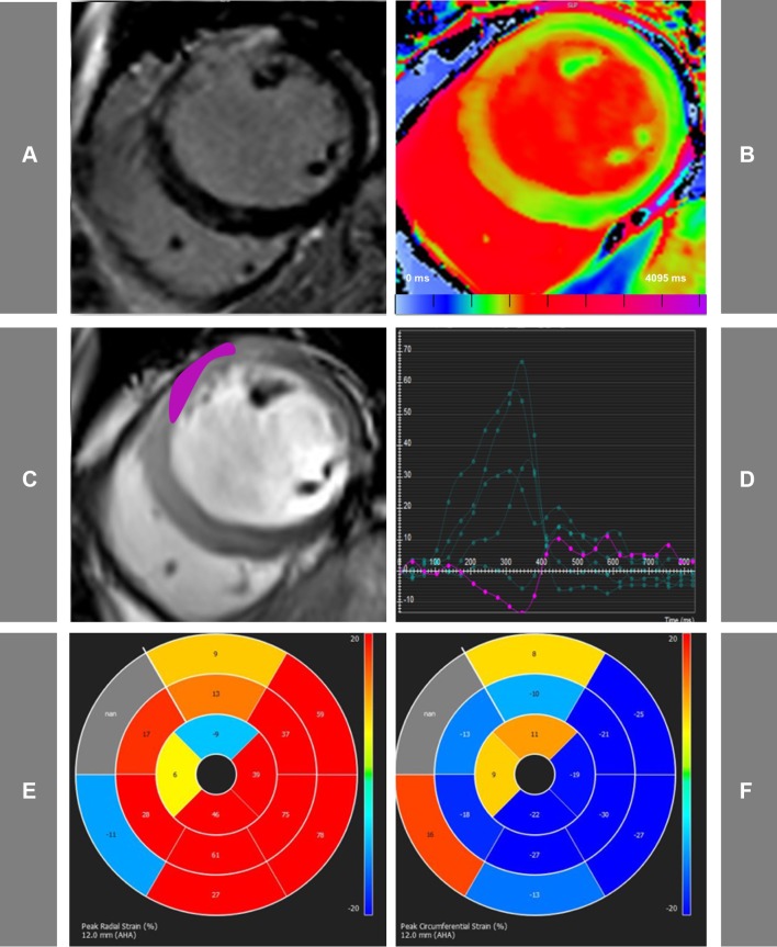
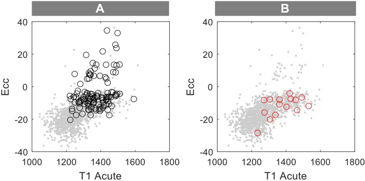
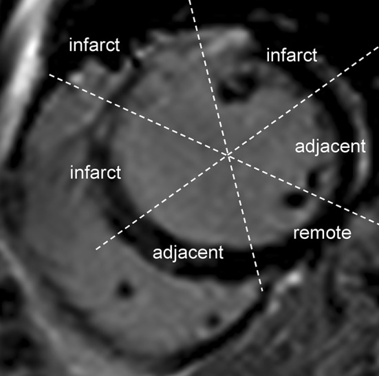
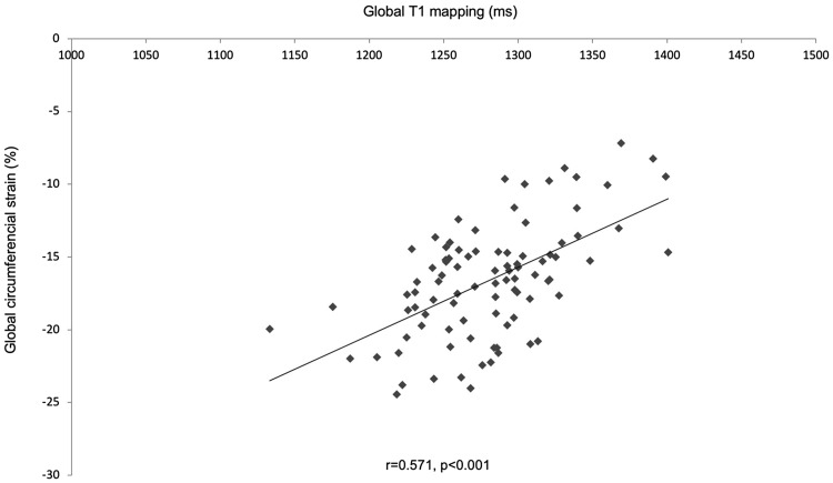
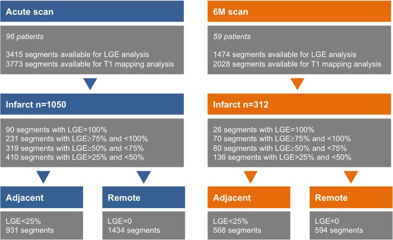
Early risk stratification after ST-segment-elevation myocardial infarction (STEMI) is of major clinical importance. Strain quantifies myocardial deformation and can demonstrate abnormal global and segmental myocardial function in acute ischaemia. Native T1-mapping allows assessment of the severity of acute ischemic injury, however its clinical applicability early post MI is limited by the complex dynamic changes happening in the myocardium post MI. We aimed to explore relationship between T1-mapping and feature tracking imaging, to establish whether combined analysis of these parameters could predict recovery after STEMI. 96 STEMI patients (aged 60 ± 11) prospectively recruited in the Oxford Acute Myocardial Infarction (OxAMI) study underwent 3T-CMR scans acutely (within 53 ± 32 h from primary percutaneous coronary intervention) and at 6 months (6M). The imaging protocol included: cine, ShMOLLI T1-mapping and late gadolinium enhancement (LGE). Segments were divided in the infarct, adjacent and remote zones based on the presence of LGE. Peak circumferential (Ecc) and radial (Err) strain was assessed using cvi42 software. Acute segmental strain correlated with segmental T1-mapping values (T1 vs. Err - 0.75 ± 0.25, p < 0.01; T1 vs. Ecc 0.72 ± 0.32, p < 0.01) and with LGE segmental injury (LGE vs. Err - 0.56 ± 0.29, p < 0.01; LGE vs. Ecc 0.54 ± 0.35, p < 0.01). Moreover, acute segmental T1 and strain predicted segmental LGE transmurality on 6M scans (p < 0.001, r = 0.5). Multiple regression analysis confirmed combined analysis of global Ecc and T1-mapping was significantly better than either method alone in predicting final infarct size at 6M (r = 0.556 vs r = 0.473 for global T1 only and r = 0.476 for global Ecc only, p < 0.001). This novel CMR method combining T1-mapping and feature tracking analysis of acute CMR scans predicts LGE transmurality and infarct size at 6M following STEMI.

摘要

ST段抬高型心肌梗死(STEMI)后的早期风险分层具有重要的临床意义。应变可量化心肌变形,并能在急性缺血时显示整体和节段性心肌功能异常。心肌组织T1加权成像可评估急性缺血损伤的严重程度,然而其在心肌梗死后早期的临床应用受到心肌梗死后心肌复杂动态变化的限制。我们旨在探讨T1加权成像与特征追踪成像之间的关系,以确定这些参数的联合分析是否能够预测STEMI后的恢复情况。在牛津急性心肌梗死(OxAMI)研究中前瞻性招募的96例STEMI患者(年龄60±11岁)在急性期(在直接经皮冠状动脉介入治疗后53±32小时内)和6个月时接受了3T心脏磁共振成像(CMR)扫描。成像方案包括:电影成像、ShMOLLI T1加权成像和延迟钆增强(LGE)。根据LGE的存在将节段分为梗死区、相邻区和远隔区。使用cvi42软件评估峰值圆周应变(Ecc)和径向应变(Err)。急性节段应变与节段T1加权成像值相关(T1与Err -0.75±0.25,p<0.01;T1与Ecc 0.72±0.32,p<0.01),并与LGE节段损伤相关(LGE与Err -0.56±0.29,p<0.01;LGE与Ecc 0.54±0.35,p<0.01)。此外,急性节段T1加权成像和应变可预测6个月扫描时节段LGE透壁性(p<0.001,r=0.5)。多元回归分析证实,在预测6个月时的最终梗死面积方面,整体Ecc和T1加权成像的联合分析明显优于单独使用任何一种方法(整体T1单独分析时r=0.556,整体Ecc单独分析时r=0.473,仅整体Ecc时r=0.476,p<0.001)。这种将T1加权成像与急性CMR扫描的特征追踪分析相结合的新型CMR方法可预测STEMI后6个月时的LGE透壁性和梗死面积。

https://cdn.ncbi.nlm.nih.gov/pmc/blobs/1cfc/6598944/197d8dc062b1/10554_2019_1542_Fig5_HTML.jpg
https://cdn.ncbi.nlm.nih.gov/pmc/blobs/1cfc/6598944/d2ab3f7e04f8/10554_2019_1542_Fig1_HTML.jpg
https://cdn.ncbi.nlm.nih.gov/pmc/blobs/1cfc/6598944/ff07f553d7e5/10554_2019_1542_Fig2_HTML.jpg
https://cdn.ncbi.nlm.nih.gov/pmc/blobs/1cfc/6598944/46132bf87d94/10554_2019_1542_Fig3_HTML.jpg
https://cdn.ncbi.nlm.nih.gov/pmc/blobs/1cfc/6598944/ca44be7d5b18/10554_2019_1542_Fig4_HTML.jpg
https://cdn.ncbi.nlm.nih.gov/pmc/blobs/1cfc/6598944/197d8dc062b1/10554_2019_1542_Fig5_HTML.jpg

相似文献

1
Combined T1-mapping and tissue tracking analysis predicts severity of ischemic injury following acute STEMI-an Oxford Acute Myocardial Infarction (OxAMI) study.联合T1映射和组织追踪分析预测急性ST段抬高型心肌梗死(STEMI)后缺血性损伤的严重程度——牛津急性心肌梗死(OxAMI)研究
Int J Cardiovasc Imaging. 2019 Jul;35(7):1297-1308. doi: 10.1007/s10554-019-01542-8. Epub 2019 Feb 16.
2
Hyper-acute cardiovascular magnetic resonance T1 mapping predicts infarct characteristics in patients with ST elevation myocardial infarction.超急性心血管磁共振 T1 映射预测 ST 段抬高型心肌梗死患者的梗死特征。
J Cardiovasc Magn Reson. 2020 Jan 9;22(1):3. doi: 10.1186/s12968-019-0593-9.
3
Cardiovascular magnetic resonance by non contrast T1-mapping allows assessment of severity of injury in acute myocardial infarction.非对比 T1 映射的心血管磁共振可评估急性心肌梗死损伤的严重程度。
J Cardiovasc Magn Reson. 2012 Feb 6;14(1):15. doi: 10.1186/1532-429X-14-15.
4
Quantification of both the area-at-risk and acute myocardial infarct size in ST-segment elevation myocardial infarction using T1-mapping.使用T1映射技术对ST段抬高型心肌梗死的梗死相关心肌面积和急性心肌梗死面积进行定量分析。
J Cardiovasc Magn Reson. 2017 Aug 1;19(1):57. doi: 10.1186/s12968-017-0370-6.
5
Acute Microvascular Impairment Post-Reperfused STEMI Is Reversible and Has Additional Clinical Predictive Value: A CMR OxAMI Study.急性再灌注后 STEMI 的微血管功能障碍是可逆的,并具有额外的临床预测价值:一项 CMR OxAMI 研究。
JACC Cardiovasc Imaging. 2019 Sep;12(9):1783-1793. doi: 10.1016/j.jcmg.2018.10.028. Epub 2019 Jan 16.
6
CMR Native T1 Mapping Allows Differentiation of Reversible Versus Irreversible Myocardial Damage in ST-Segment-Elevation Myocardial Infarction: An OxAMI Study (Oxford Acute Myocardial Infarction).心脏磁共振成像(CMR)的固有T1映射可区分ST段抬高型心肌梗死中可逆性与不可逆性心肌损伤:一项牛津急性心肌梗死(OxAMI)研究
Circ Cardiovasc Imaging. 2017 Aug;10(8):e005986. doi: 10.1161/CIRCIMAGING.116.005986.
7
Myocardial Extracellular Volume Estimation by CMR Predicts Functional Recovery Following Acute MI.CMR 估算心肌细胞外容积可预测急性心肌梗死后的功能恢复。
JACC Cardiovasc Imaging. 2017 Sep;10(9):989-999. doi: 10.1016/j.jcmg.2016.06.015. Epub 2016 Oct 19.
8
Relationship of Myocardial Strain and Markers of Myocardial Injury to Predict Segmental Recovery After Acute ST-Segment-Elevation Myocardial Infarction.急性ST段抬高型心肌梗死心肌应变与心肌损伤标志物的关系对节段恢复的预测作用
Circ Cardiovasc Imaging. 2016 Jun;9(6). doi: 10.1161/CIRCIMAGING.115.003457.
9
Novel insights into myocardial fibrosis in patients with new onset ST-elevation myocardial infarction following percutaneous coronary intervention through enhanced cardiac magnetic resonance imaging: a prospective cohort study.通过增强心脏磁共振成像对经皮冠状动脉介入治疗后新发ST段抬高型心肌梗死患者心肌纤维化的新见解:一项前瞻性队列研究。
BMC Cardiovasc Disord. 2025 Apr 10;25(1):274. doi: 10.1186/s12872-025-04719-3.
10
Dynamic changes in injured myocardium, very early after acute myocardial infarction, quantified using T1 mapping cardiovascular magnetic resonance.采用 T1 mapping 心血管磁共振技术定量检测急性心肌梗死后极早期损伤心肌的动态变化。
J Cardiovasc Magn Reson. 2018 Dec 20;20(1):82. doi: 10.1186/s12968-018-0506-3.

引用本文的文献

1
Role of stroke volume in patients presenting with uncomplicated anterior STEMI.每搏输出量在无并发症的前壁ST段抬高型心肌梗死患者中的作用。
Open Heart. 2025 Sep 5;12(2):e003429. doi: 10.1136/openhrt-2025-003429.
2
Prognostic Value of Strain by Tissue Tracking Cardiac Magnetic Resonance in Myocardial Infarction With Nonobstructive Coronary Arteries.组织追踪心脏磁共振成像测量应变在非阻塞性冠状动脉心肌梗死中的预后价值
J Am Heart Assoc. 2025 Apr 15;14(8):e039395. doi: 10.1161/JAHA.124.039395. Epub 2025 Apr 7.
3
Clinical Applications of Cardiac Magnetic Resonance Parametric Mapping.

本文引用的文献

1
Cardiac Magnetic Resonance Myocardial Feature Tracking for Optimized Prediction of Cardiovascular Events Following Myocardial Infarction.心肌磁共振心肌特征追踪优化预测心肌梗死后心血管事件。
JACC Cardiovasc Imaging. 2018 Oct;11(10):1433-1444. doi: 10.1016/j.jcmg.2017.11.034. Epub 2018 Feb 14.
2
Acute Infarct Extracellular Volume Mapping to Quantify Myocardial Area at Risk and Chronic Infarct Size on Cardiovascular Magnetic Resonance Imaging.急性梗死细胞外容积映射以量化心血管磁共振成像上的心肌危险区和慢性梗死大小。
Circ Cardiovasc Imaging. 2017 Jul;10(7). doi: 10.1161/CIRCIMAGING.117.006182.
3
Prognostic Significance of Remote Myocardium Alterations Assessed by Quantitative Noncontrast T1 Mapping in ST-Segment Elevation Myocardial Infarction.
心脏磁共振参数成像的临床应用
Diagnostics (Basel). 2024 Aug 20;14(16):1816. doi: 10.3390/diagnostics14161816.
4
Prognostic value of visual and quantitative CMR regional myocardial function in patients with suspected myocarditis.疑似心肌炎患者的视觉和定量 CMR 区域性心肌功能的预后价值。
Int J Cardiovasc Imaging. 2024 Apr;40(4):907-920. doi: 10.1007/s10554-024-03059-1. Epub 2024 Mar 1.
5
Microvascular Obstruction in Acute Myocardial Infarction, a Potential Therapeutic Target.急性心肌梗死中的微血管阻塞:一个潜在的治疗靶点
J Clin Med. 2023 Sep 13;12(18):5934. doi: 10.3390/jcm12185934.
6
Comparison of manual and artificial intelligence based quantification of myocardial strain by feature tracking-a cardiovascular MR study in health and disease.基于特征追踪的心肌应变手动和人工智能定量比较——健康和疾病的心血管磁共振研究。
Eur Radiol. 2024 Feb;34(2):1003-1015. doi: 10.1007/s00330-023-10127-y. Epub 2023 Aug 18.
7
Prospects for Precision Medicine in Acute Myocardial Infarction: Patient-Level Insights into Myocardial Injury and Repair.急性心肌梗死精准医学的前景:关于心肌损伤与修复的患者层面见解
J Clin Med. 2023 Jul 13;12(14):4668. doi: 10.3390/jcm12144668.
8
Artificial Intelligence as a Diagnostic Tool in Non-Invasive Imaging in the Assessment of Coronary Artery Disease.人工智能作为一种诊断工具在非侵入性成像评估冠状动脉疾病中的应用。
Med Sci (Basel). 2023 Feb 24;11(1):20. doi: 10.3390/medsci11010020.
9
Patient-Specific Cardiac Magnetic Resonance Feature Tracking Approach for Scar Detection in Concomitant Ischemic and Non-Ischemic Heart Disease.用于同时存在缺血性和非缺血性心脏病时瘢痕检测的患者特异性心脏磁共振特征跟踪方法
Cardiol Cardiovasc Med. 2022;6(6):542-549. doi: 10.26502/fccm.92920297. Epub 2022 Dec 23.
10
Elevated Native T1 Values in the Remote Myocardium Supplied by Obstructive Non-Infarct Related Coronary Arteries in Post-STEMI Cardiac Magnetic Resonance.梗死后心肌磁共振中阻塞性非梗死相关冠状动脉供应的远段心肌 T1 值升高。
Cardiology. 2023;148(2):106-113. doi: 10.1159/000528143. Epub 2022 Nov 22.
定量对比增强 T1 mapping 技术评估 ST 段抬高型心肌梗死患者远隔心肌改变的预后意义
JACC Cardiovasc Imaging. 2018 Mar;11(3):411-419. doi: 10.1016/j.jcmg.2017.03.015. Epub 2017 Jun 14.
4
Effects of irrelevant background speech on eye movements during reading.无关背景语音对阅读过程中眼动的影响。
Q J Exp Psychol (Hove). 2018 Jun;71(6):1270-1275. doi: 10.1080/17470218.2017.1339718. Epub 2018 Jan 1.
5
Cardiac and Noncardiac Causes of Long-Term Mortality in ST-Segment-Elevation Acute Myocardial Infarction Patients Who Underwent Primary Percutaneous Coronary Intervention.接受直接经皮冠状动脉介入治疗的ST段抬高型急性心肌梗死患者长期死亡的心脏和非心脏原因
Circ Cardiovasc Qual Outcomes. 2017 Jan;10(1). doi: 10.1161/CIRCOUTCOMES.116.002790.
6
Myocardial Extracellular Volume Estimation by CMR Predicts Functional Recovery Following Acute MI.CMR 估算心肌细胞外容积可预测急性心肌梗死后的功能恢复。
JACC Cardiovasc Imaging. 2017 Sep;10(9):989-999. doi: 10.1016/j.jcmg.2016.06.015. Epub 2016 Oct 19.
7
From Recognized to Novel Quantitative CMR Biomarkers of LV Recovery: A Paradigm Shift in Acute Myocardial Infarction Imaging.从公认的左心室恢复定量CMR生物标志物到新型标志物:急性心肌梗死成像的范式转变
JACC Cardiovasc Imaging. 2017 Sep;10(9):1000-1002. doi: 10.1016/j.jcmg.2016.07.007. Epub 2016 Oct 19.
8
Early Gadolinium Enhancement for Determination of Area at Risk: A Preclinical Validation Study.早期钆增强用于确定危险区域:一项临床前验证研究。
JACC Cardiovasc Imaging. 2017 Feb;10(2):130-139. doi: 10.1016/j.jcmg.2016.04.009. Epub 2016 Sep 21.
9
Remote Zone Extracellular Volume and Left Ventricular Remodeling in Survivors of ST-Elevation Myocardial Infarction.ST段抬高型心肌梗死幸存者的远隔区细胞外容积与左心室重构
Hypertension. 2016 Aug;68(2):385-91. doi: 10.1161/HYPERTENSIONAHA.116.07222. Epub 2016 Jun 27.
10
Temporal Evolution of Myocardial Hemorrhage and Edema in Patients After Acute ST-Segment Elevation Myocardial Infarction: Pathophysiological Insights and Clinical Implications.急性ST段抬高型心肌梗死后患者心肌出血和水肿的时间演变:病理生理见解及临床意义
J Am Heart Assoc. 2016 Feb 23;5(2):e002834. doi: 10.1161/JAHA.115.002834.