• 文献检索
  • 文档翻译
  • 深度研究
  • 学术资讯
  • Suppr Zotero 插件Zotero 插件
  • 邀请有礼
  • 套餐&价格
  • 历史记录
应用&插件
Suppr Zotero 插件Zotero 插件浏览器插件Mac 客户端Windows 客户端微信小程序
定价
高级版会员购买积分包购买API积分包
服务
文献检索文档翻译深度研究API 文档MCP 服务
关于我们
关于 Suppr公司介绍联系我们用户协议隐私条款
关注我们

Suppr 超能文献

核心技术专利:CN118964589B侵权必究
粤ICP备2023148730 号-1Suppr @ 2026

文献检索

告别复杂PubMed语法,用中文像聊天一样搜索,搜遍4000万医学文献。AI智能推荐,让科研检索更轻松。

立即免费搜索

文件翻译

保留排版,准确专业,支持PDF/Word/PPT等文件格式,支持 12+语言互译。

免费翻译文档

深度研究

AI帮你快速写综述,25分钟生成高质量综述,智能提取关键信息,辅助科研写作。

立即免费体验

通过增强心脏磁共振成像对经皮冠状动脉介入治疗后新发ST段抬高型心肌梗死患者心肌纤维化的新见解:一项前瞻性队列研究。

Novel insights into myocardial fibrosis in patients with new onset ST-elevation myocardial infarction following percutaneous coronary intervention through enhanced cardiac magnetic resonance imaging: a prospective cohort study.

作者信息

Oketunbi Temilola J, Wang Jun, Ding Bin, Song Xilong, Li Yao, Song Hongwei, Shi Xiaojun, Hu Sigang, Gao Dasheng, Wang Hongju, Li Miaonan

机构信息

Department of Cardiovascular Diseases, The First Affiliated Hospital of Bengbu Medical University, Bengbu, 233004, China.

Department of Radiology, The First Affiliated Hospital of Bengbu Medical University, Bengbu, 233004, China.

出版信息

BMC Cardiovasc Disord. 2025 Apr 10;25(1):274. doi: 10.1186/s12872-025-04719-3.

DOI:10.1186/s12872-025-04719-3
PMID:40211110
原文链接:https://pmc.ncbi.nlm.nih.gov/articles/PMC11983772/
Abstract

BACKGROUND

Myocardial fibrosis is a prevalent pathological hallmark of a diverse range of chronic and acute cardiovascular disorders. However, the relevant literature currently provides limited evidence regarding the determinants of myocardial fibrosis severity in patients with new-onset ST-elevation myocardial infarction (STEMI) following successful emergent percutaneous coronary intervention (PCI) utilizing contrast-enhanced cardiac magnetic resonance imaging (CE-CMR).

METHODS

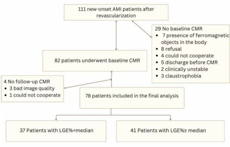
We prospectively enrolled a cohort of 78 patients who presented with new-onset ST-segment elevation myocardial infarction and who underwent successful emergent PCI within 12 h from the onset of symptoms. Late gadolinium-enhanced LGE (LGE) was quantified via CE-CMR, and patients were categorized into two groups on the basis of the median LGE value.

RESULTS

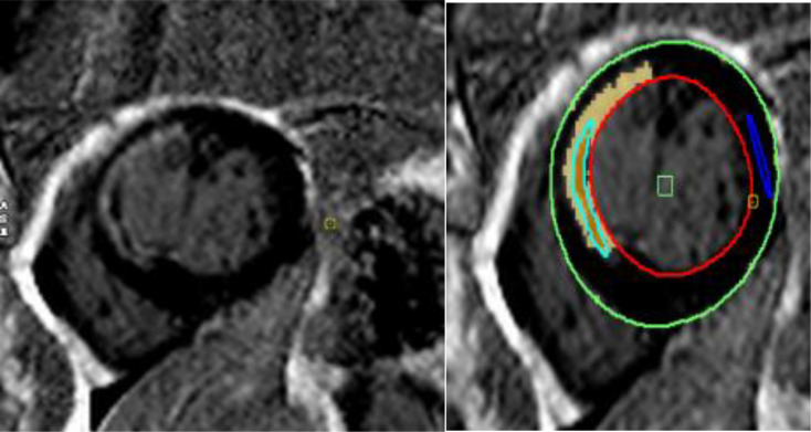
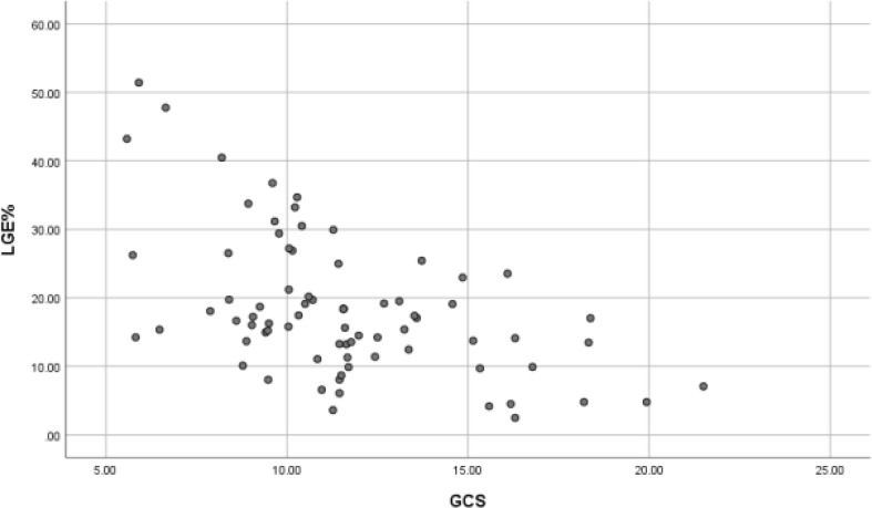
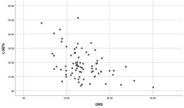
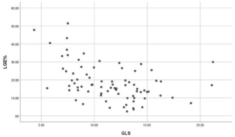
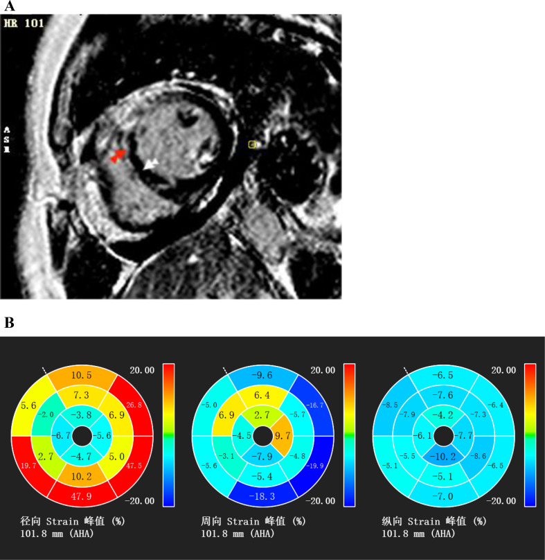
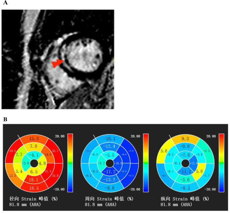
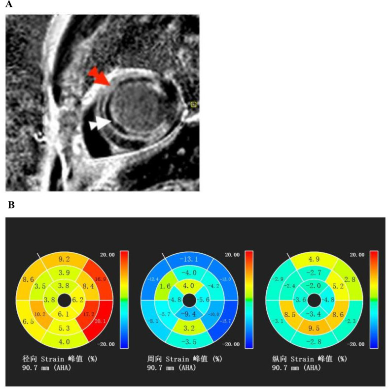
The median LGE was 16% (IQR 12 to 24). Compared with patients with LGE below the median (n = 37), those with LGE above the median (n = 41) presented significantly reduced left ventricular global radial strain(GRS), global circumferential strain(GCS), and global longitudinal strain(GLS) (all p < 0.05). The infarcted radial segment (IRS), infarcted circumferential segment (ICS) and infarcted longitudinal segment (ILS) were significantly reduced in patients with greater LGE (all p < 0.05). The occurrence rates of microvascular obstruction (MVO) (p < 0.001) and wall motion abnormality (WMA) (p < 0.01) were significantly greater in patients with a greater extent of LGE, despite successful reperfusion therapy. LGE exhibited a moderate negative correlation with the global circumferential segment (r=-0.547, p < 0.001) and a weak negative correlation with both the global radial segment and the global longitudinal segment (r=-0.434, p < 0.001; r=-0.437, p < 0.001). In the multivariable linear regression analysis model, the Gensini score (β = 0.258; p < 0.01), LVEF% (β=-0.269; p < 0.05), MVO (β = 0.343; p < 0.001) and GRS (β = 0.227; p < 0.05) emerged as robust predictors of myocardial fibrosis.

CONCLUSION

The present study revealed a correlation of cardiac pathological structure, microcirculation, and myocardial fibrosis in the context of acute myocardial infarction. Therefore, this study provides theoretical evidence from a pathological perspective regarding the progression of myocardial fibrosis in patients with new-onset STEMI following successful PCI.

TRIAL REGISTRATION

The trial was registered in the Chinese Clinical Trial Registry (ChiCTR2400080282; January 25th, 2024).

摘要

背景

心肌纤维化是多种急慢性心血管疾病普遍存在的病理特征。然而,目前相关文献中关于采用对比增强心脏磁共振成像(CE-CMR)对首次发生ST段抬高型心肌梗死(STEMI)患者成功进行急诊经皮冠状动脉介入治疗(PCI)后心肌纤维化严重程度的决定因素的证据有限。

方法

我们前瞻性纳入了一组78例首次发生ST段抬高型心肌梗死且在症状发作后12小时内成功进行急诊PCI的患者。通过CE-CMR对延迟钆增强(LGE)进行定量分析,并根据LGE值的中位数将患者分为两组。

结果

LGE的中位数为16%(四分位间距12%至24%)。与LGE低于中位数的患者(n = 37)相比,LGE高于中位数的患者(n = 41)左心室整体径向应变(GRS)、整体圆周应变(GCS)和整体纵向应变(GLS)均显著降低(均p < 0.05)。LGE较高的患者梗死径向节段(IRS)、梗死圆周节段(ICS)和梗死纵向节段(ILS)均显著减少(均p < 0.05)。尽管进行了成功的再灌注治疗,但LGE程度较高的患者微血管阻塞(MVO)(p < 0.001)和室壁运动异常(WMA)(p < 0.01)的发生率显著更高。LGE与整体圆周节段呈中度负相关(r = -0.547,p < 0.001),与整体径向节段和整体纵向节段均呈弱负相关(r = -0.434,p < 0.001;r = -0.437,p < 0.001)。在多变量线性回归分析模型中,Gensini评分(β = 0.258;p < 0.01)、左心室射血分数百分比(LVEF%)(β = -0.269;p < 0.05)、MVO(β = 0.343;p < 0.001)和GRS(β = 0.227;p < 0.05)是心肌纤维化的有力预测指标。

结论

本研究揭示了急性心肌梗死背景下心脏病理结构、微循环与心肌纤维化之间的相关性。因此,本研究从病理角度为首次发生STEMI患者成功PCI后心肌纤维化的进展提供了理论依据。

试验注册

该试验已在中国临床试验注册中心注册(ChiCTR2400080282;2024年1月25日)。

https://cdn.ncbi.nlm.nih.gov/pmc/blobs/b78c/11983772/15f2eca5a842/12872_2025_4719_Fig8_HTML.jpg
https://cdn.ncbi.nlm.nih.gov/pmc/blobs/b78c/11983772/9d73757b8f58/12872_2025_4719_Fig1_HTML.jpg
https://cdn.ncbi.nlm.nih.gov/pmc/blobs/b78c/11983772/976297a188fd/12872_2025_4719_Fig2_HTML.jpg
https://cdn.ncbi.nlm.nih.gov/pmc/blobs/b78c/11983772/47f1ea449aba/12872_2025_4719_Fig3_HTML.jpg
https://cdn.ncbi.nlm.nih.gov/pmc/blobs/b78c/11983772/d86efc10ec89/12872_2025_4719_Fig4_HTML.jpg
https://cdn.ncbi.nlm.nih.gov/pmc/blobs/b78c/11983772/2b8b5d886824/12872_2025_4719_Fig5_HTML.jpg
https://cdn.ncbi.nlm.nih.gov/pmc/blobs/b78c/11983772/41321c940e5c/12872_2025_4719_Fig6_HTML.jpg
https://cdn.ncbi.nlm.nih.gov/pmc/blobs/b78c/11983772/143955615a12/12872_2025_4719_Fig7_HTML.jpg
https://cdn.ncbi.nlm.nih.gov/pmc/blobs/b78c/11983772/15f2eca5a842/12872_2025_4719_Fig8_HTML.jpg

相似文献

1
Novel insights into myocardial fibrosis in patients with new onset ST-elevation myocardial infarction following percutaneous coronary intervention through enhanced cardiac magnetic resonance imaging: a prospective cohort study.通过增强心脏磁共振成像对经皮冠状动脉介入治疗后新发ST段抬高型心肌梗死患者心肌纤维化的新见解:一项前瞻性队列研究。
BMC Cardiovasc Disord. 2025 Apr 10;25(1):274. doi: 10.1186/s12872-025-04719-3.
2
Association of stress hyperglycemia ratio with left ventricular function and microvascular obstruction in patients with ST-segment elevation myocardial infarction: a 3.0 T cardiac magnetic resonance study.应激性高血糖比值与 ST 段抬高型心肌梗死患者左心室功能和微血管阻塞的关系:一项 3.0T 心脏磁共振研究。
Cardiovasc Diabetol. 2024 May 27;23(1):179. doi: 10.1186/s12933-024-02271-6.
3
Hyper-acute cardiovascular magnetic resonance T1 mapping predicts infarct characteristics in patients with ST elevation myocardial infarction.超急性心血管磁共振 T1 映射预测 ST 段抬高型心肌梗死患者的梗死特征。
J Cardiovasc Magn Reson. 2020 Jan 9;22(1):3. doi: 10.1186/s12968-019-0593-9.
4
Predictive value of myocardial strain on myocardial infarction size by cardiac magnetic resonance imaging in ST-segment elevation myocardial infarction with preserved left ventricular ejection fraction.心脏磁共振成像评估心肌应变对左心室射血分数保留的ST段抬高型心肌梗死患者心肌梗死面积的预测价值
Front Pharmacol. 2022 Oct 14;13:1015390. doi: 10.3389/fphar.2022.1015390. eCollection 2022.
5
[Predictive value of global longitudinal strain measured by cardiac magnetic resonance imaging for left ventricular remodeling after acute ST-segment elevation myocardial infarction: a multi-centered prospective study].[心脏磁共振成像测量的整体纵向应变对急性ST段抬高型心肌梗死左心室重构的预测价值:一项多中心前瞻性研究]
Nan Fang Yi Ke Da Xue Xue Bao. 2024 Jun 20;44(6):1033-1039. doi: 10.12122/j.issn.1673-4254.2024.06.03.
6
Combined T1-mapping and tissue tracking analysis predicts severity of ischemic injury following acute STEMI-an Oxford Acute Myocardial Infarction (OxAMI) study.联合T1映射和组织追踪分析预测急性ST段抬高型心肌梗死(STEMI)后缺血性损伤的严重程度——牛津急性心肌梗死(OxAMI)研究
Int J Cardiovasc Imaging. 2019 Jul;35(7):1297-1308. doi: 10.1007/s10554-019-01542-8. Epub 2019 Feb 16.
7
The role of left ventricular deformation in the assessment of microvascular obstruction and intramyocardial haemorrhage.左心室变形在评估微血管阻塞和心肌内出血中的作用。
Int J Cardiovasc Imaging. 2017 Mar;33(3):361-370. doi: 10.1007/s10554-016-1006-x. Epub 2016 Oct 26.
8
Association Between Haptoglobin Phenotype and Microvascular Obstruction in Patients With STEMI: A Cardiac Magnetic Resonance Study.载脂蛋白表型与 STEMI 患者微血管阻塞的相关性:一项心脏磁共振研究。
JACC Cardiovasc Imaging. 2019 Jun;12(6):1007-1017. doi: 10.1016/j.jcmg.2018.03.004. Epub 2018 Apr 18.
9
Myocardial Extracellular Volume Estimation by CMR Predicts Functional Recovery Following Acute MI.CMR 估算心肌细胞外容积可预测急性心肌梗死后的功能恢复。
JACC Cardiovasc Imaging. 2017 Sep;10(9):989-999. doi: 10.1016/j.jcmg.2016.06.015. Epub 2016 Oct 19.
10
[Predictive value of cardiac magnetic resonance imaging for adverse left ventricular remodeling after acute ST-segment elevation myocardial infarction].[心脏磁共振成像对急性ST段抬高型心肌梗死左心室不良重构的预测价值]
Nan Fang Yi Ke Da Xue Xue Bao. 2024 Mar 20;44(3):553-562. doi: 10.12122/j.issn.1673-4254.2024.03.17.

本文引用的文献

1
Cardiac magnetic resonance imaging-derived pathophysiology and prognosis of diabetes mellitus with acute myocardial infarction after revascularization: a prospective cohort study.经心脏磁共振成像评估的糖尿病合并经血运重建治疗的急性心肌梗死后的病理生理学和预后:一项前瞻性队列研究。
Ann Med. 2024 Dec;56(1):2399751. doi: 10.1080/07853890.2024.2399751. Epub 2024 Sep 10.
2
Multimodal data-driven prognostic model for predicting new-onset ST-elevation myocardial infarction following emergency percutaneous coronary intervention.用于预测急诊经皮冠状动脉介入治疗后新发ST段抬高型心肌梗死的多模态数据驱动预后模型。
Inflamm Res. 2023 Sep;72(9):1799-1809. doi: 10.1007/s00011-023-01781-5. Epub 2023 Aug 29.
3
Predicting long-term prognosis after percutaneous coronary intervention in patients with new onset ST-elevation myocardial infarction: development and external validation of a nomogram model.
预测新发 ST 段抬高型心肌梗死患者经皮冠状动脉介入治疗后的长期预后:列线图模型的建立与外部验证。
Cardiovasc Diabetol. 2023 Apr 13;22(1):87. doi: 10.1186/s12933-023-01820-9.
4
Comprehensive Phenotypic Characterization of Late Gadolinium Enhancement Predicts Sudden Cardiac Death in Coronary Artery Disease.全面表型特征分析预测冠状动脉疾病患者的心脏性猝死。
JACC Cardiovasc Imaging. 2023 May;16(5):628-638. doi: 10.1016/j.jcmg.2022.10.020. Epub 2023 Jan 11.
5
Cardiac magnetic resonance shows increased adverse ventricular remodeling in younger patients after ST-segment elevation myocardial infarction.心脏磁共振显示 ST 段抬高型心肌梗死患者中年轻患者的不良心室重构增加。
Eur Radiol. 2023 Jul;33(7):4637-4647. doi: 10.1007/s00330-023-09406-5. Epub 2023 Jan 26.
6
Plaque histology and myocardial disease in sudden coronary death: the Fingesture study.斑块组织病理学和冠状动脉猝死中的心肌疾病:Fingesture 研究。
Eur Heart J. 2022 Dec 14;43(47):4923-4930. doi: 10.1093/eurheartj/ehac533.
7
Deceleration Capacity Improves Prognostic Accuracy of Relative Increase and Final Coronary Physiology in Patients With Non-ST-Elevation Acute Coronary Syndrome.减速能力提高非ST段抬高型急性冠脉综合征患者相对增加和最终冠脉生理学的预后准确性。
Front Cardiovasc Med. 2022 Mar 22;9:848499. doi: 10.3389/fcvm.2022.848499. eCollection 2022.
8
Diagnosis and Treatment of Acute Coronary Syndromes: A Review.急性冠状动脉综合征的诊断与治疗:综述。
JAMA. 2022 Feb 15;327(7):662-675. doi: 10.1001/jama.2022.0358.
9
Intramyocardial Hemorrhage and the "Wave Front" of Reperfusion Injury Compromising Myocardial Salvage.心肌内出血与再灌注损伤的“波阵面”危及心肌挽救。
J Am Coll Cardiol. 2022 Jan 4;79(1):35-48. doi: 10.1016/j.jacc.2021.10.034.
10
Functional and prognostic implications of cardiac magnetic resonance feature tracking-derived remote myocardial strain analyses in patients following acute myocardial infarction.心脏磁共振特征追踪衍生的远程心肌应变分析在急性心肌梗死后患者中的功能和预后意义。
Clin Res Cardiol. 2021 Feb;110(2):270-280. doi: 10.1007/s00392-020-01747-1. Epub 2020 Oct 20.