California Institute of Technology, Caltech Optical Imaging Laboratory, Department of Electrical Eng, United States.
California Institute of Technology, Caltech Optical Imaging Laboratory, Andrew and Peggy Cheng Depar, United States.
J Biomed Opt. 2019 Feb;24(2):1-8. doi: 10.1117/1.JBO.24.2.026003.
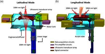
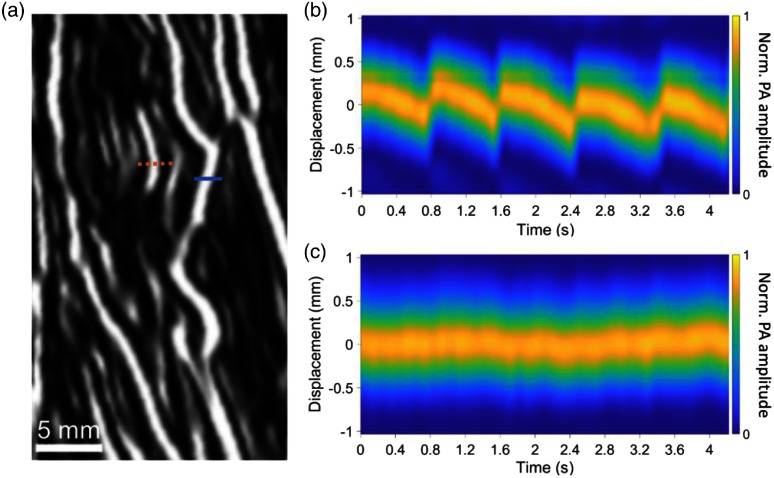
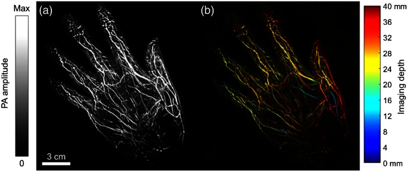
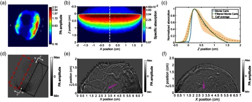
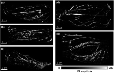
We present a method of imaging angiographic structures in human extremities, including hands, arms, legs, and feet, using a newly developed photoacoustic computed tomography (PACT) system. The system features deep penetration (1.8 cm in muscular tissues) with high spatial and temporal resolutions. A volumetric image is acquired within 5 to 15 s while each cross sectional image is acquired within 100 μs. Therefore, we see no blurring from motion in the imaging plane. Longitudinal and latitudinal cross-sectional images of a healthy volunteer clearly show the vascular network of each appendage and highlight the system's ability to image major and minor vasculatures, without the use of an external contrast or ionizing radiation. We also track heartbeat-induced arterial movement at a two-dimensional frame rate of 10 Hz. This work substantiates the idea that PACT could be used as a noninvasive method for imaging human vasculatures.
我们提出了一种使用新开发的光声计算机断层扫描(PACT)系统对人体四肢(包括手、臂、腿和脚)的血管结构进行成像的方法。该系统具有深穿透性(在肌肉组织中为 1.8 厘米)和高空间和时间分辨率。在 5 到 15 秒内获取体积图像,而在 100 微秒内获取每个横截面图像。因此,我们在成像平面上没有看到运动模糊。健康志愿者的纵向和横向横截面图像清楚地显示了每个附属物的血管网络,并突出了该系统无需使用外部对比剂或电离辐射即可成像主要和次要血管的能力。我们还以 10 Hz 的二维帧率跟踪心跳引起的动脉运动。这项工作证实了 PACT 可作为一种非侵入性方法用于人体血管成像的想法。