University of Cambridge Metabolic Research Laboratories and MRC Metabolic Diseases Unit, Wellcome Trust-MRC Institute of Metabolic Science, Addenbrooke's Treatment Centre, Addenbrooke's Hospital, Cambridge, UK.
University Department of Obstetrics and Gynaecology, University of Cambridge, Cambridge, UK.
J Physiol. 2019 May;597(9):2391-2401. doi: 10.1113/JP277431. Epub 2019 Mar 24.
Exposure to chronic hypoxia during gestation influences long-term health and development, including reproductive capacity, across generations. If the peri-conceptual environment in the developing oviduct is affected by gestational hypoxia, then this could have implications for later fertility and the health of future generations. In the present study, we show that the oviducts of female rats exposed to chronic hypoxia in utero have reduced telomere length, decreased mitochondrial DNA biogenesis and increased oxidative stress The results of the present study show that exposure to chronic gestational hypoxia leads to accelerated ageing of the oviduct in early adulthood and they help us understand how exposure to hypoxia during development could influence reproductive health across generations.
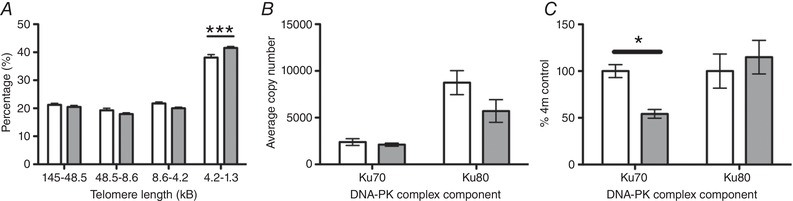
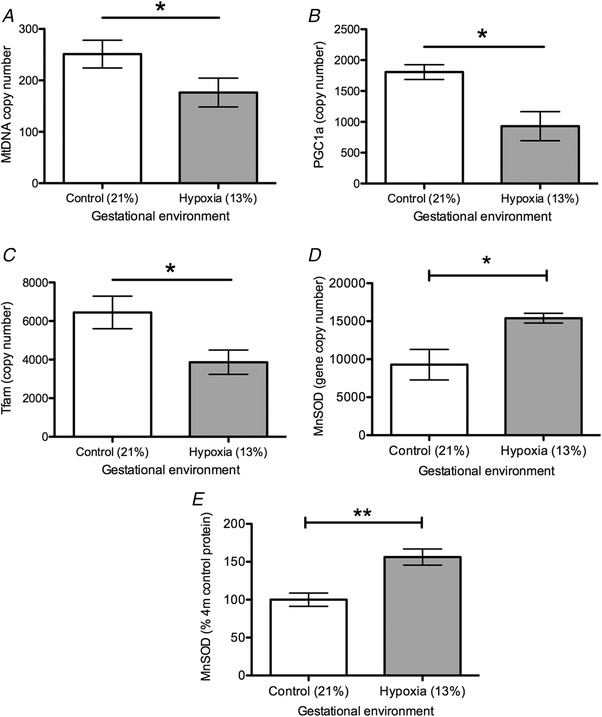
Exposure to chronic hypoxia during fetal development has important effects on immediate and long-term outcomes in offspring. Adverse impacts in adult offspring include impairment of cardiovascular function, metabolic derangement and accelerated ovarian ageing. However, it is not known whether other aspects of the female reproductive system may be similarly affected. In the present study, we examined the impact of chronic gestational hypoxia on the developing oviduct. Wistar rat dams were randomized to either normoxia (21%) or hypoxia (13%) from day 6 post-mating until delivery. Post-delivery female offspring were maintained in normoxia until 4 months of age. Oviductal gene expression was assayed at the RNA (quantitative RT-PCR) and protein (western blotting) levels. Oviductal telomere length was assayed using Southern blotting. Oviductal telomere length was reduced in the gestational hypoxia-exposed animals compared to normoxic controls (P < 0.01). This was associated with a specific post-transcriptional reduction in the KU70 subunit of DNA-pk in the gestational hypoxia-exposed group (P < 0.05). Gestational hypoxia-exposed oviducts also showed evidence of decreased mitochondrial DNA biogenesis, reduced mtDNA copy number (P < 0.05) and reduced gene expression of Tfam (P < 0.05) and Pgc1α (P < 0.05). In the hypoxia-exposed oviducts, there was upregulation of mitochondrial-specific anti-oxidant defence enzymes (MnSOD; P < 0.01). Exposure to chronic gestational hypoxia leads to accelerated ageing of the oviduct in adulthood. The oviduct plays a central role in early development as the site of gamete transport, syngamy, and early development; hence, accelerated ageing of the oviductal environment could have important implications for fertility and the health of future generations.
孕期慢性缺氧会影响长期健康和发育,包括生殖能力和代际间的生殖能力。如果发育中的输卵管的围孕期环境受到妊娠期缺氧的影响,那么这可能会对以后的生育能力和后代的健康产生影响。在本研究中,我们发现,宫内暴露于慢性缺氧的雌性大鼠的输卵管端粒长度缩短,线粒体 DNA 生物发生减少,氧化应激增加。本研究结果表明,慢性妊娠期缺氧会导致成年早期输卵管加速老化,这有助于我们了解发育过程中暴露于缺氧如何影响代际生殖健康。
胎儿发育过程中暴露于慢性缺氧对后代的即时和长期结果有重要影响。成年后代的不良影响包括心血管功能受损、代谢紊乱和卵巢衰老加速。然而,尚不清楚女性生殖系统的其他方面是否也可能受到类似的影响。在本研究中,我们研究了慢性妊娠期缺氧对发育中的输卵管的影响。Wistar 大鼠母鼠从交配后第 6 天开始随机分为常氧(21%)或缺氧(13%)组,直至分娩。产后雌性后代在常氧环境中维持至 4 月龄。使用定量 RT-PCR 和 Western 印迹法检测输卵管的基因表达。使用Southern 印迹法检测输卵管端粒长度。与正常对照组相比,宫内缺氧暴露组的输卵管端粒长度缩短(P<0.01)。这与 DNA-PK 的 KU70 亚单位在宫内缺氧暴露组的特定转录后减少有关(P<0.05)。宫内缺氧暴露的输卵管也显示出线粒体 DNA 生物发生减少、mtDNA 拷贝数减少(P<0.05)以及 Tfam(P<0.05)和 Pgc1α(P<0.05)基因表达减少的证据。在缺氧暴露的输卵管中,线粒体特异性抗氧化防御酶(MnSOD;P<0.01)上调。慢性妊娠期缺氧暴露导致成年后输卵管加速老化。输卵管在配子运输、合子形成和早期发育中起着核心作用;因此,输卵管环境的加速老化可能对生育能力和后代的健康产生重要影响。