Li Shixu, Hu Yiping, Guo Ran, Shao Yushuang, Zhao Jiangyue, Zhang Jinsong, Wang Jing
Department of Ophthalmology, the Fourth Affiliated Hospital of China Medical University, Eye Hospital of China Medical University, the Key Lenticular Laboratory of Liaoning Province, Shenyang, 110005, China.
BMC Ophthalmol. 2019 Feb 21;19(1):59. doi: 10.1186/s12886-019-1068-3.
To evaluate the effects of anterior capsular opening size on deviation from predicted refraction and the effective position of the intraocular lens (ELP) in cataract surgery.
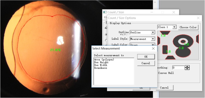
Nonrandomized clinical trial. Eighty patients (80 eyes) with simple age-related cataracts were treated from May 2018 to September 2018 at the Fourth Affiliated Hospital of China Medical University. All patients undergoing phacoemulsification received intraocular lens based on the voluntary principle. Forty eyes were implanted with the C-loop haptic intraocular lens (AMO Tecnis ZCB00) while the other 40 eyes were implanted with the plate haptic intraocular lens (CT ASPHINA 509 M). Follow-up visits were conducted postoperatively at 1 week, 1 month, and 3 months during which patients underwent refraction and data collection after pupil dilation, which included anterior segment photography and Scheimpflug imaging by Pentacam. The area, horizontal and vertical diameter of the capsulorrhexis, circularity, decentration, and package were analysed using the image analysis software Image-Pro-Plus 6.0,then evaluated the relationship between the different shapes of capsulorrhexis with deviation from predicted refraction and ELP in cataract surgery.
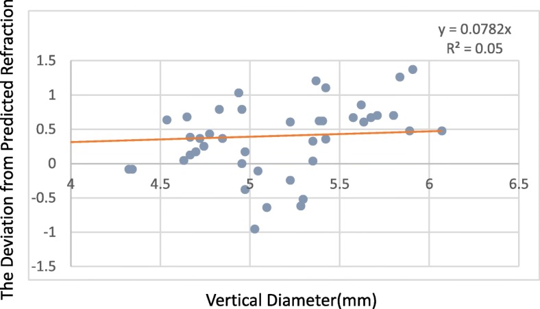
Deviation from predicted refraction and all of the parameters of capsulorrhexis were not correlative in the 509 M IOL group, however, in the Tecnis IOL group, while the deviation from predicted refraction and all of the capsulorrhexis parameters were not correlative at 1 week, the deviation from predicted refraction did correlate with capsulorrhexis area, horizontal diameter at 1 month (P = 0.029, P = 0.048), and with capsulorrhexis area, vertical diameter at 3 months (P = 0.03, P = 0.017). The ELP correlated with package in both groups postoperatively (r > 0, P < 0.05), but there is no other capsulorrhexis parameters correlated with ELP in the 509 M IOL group (all P > 0.05). For the Tecnis IOL group, the ELP and capsulorrhexis area were correlated at 1 week and 1 month, while the ELP and horizontal diameter, the ELP and vertical diameter were correlated at 1 week, but did not correlate with the other capsulorrhexis parameters in the Tecnis IOL group (all P > 0.05).
The shape of the capsulorrhexis has an effect on postoperative refractive outcomes and the effective position of the intraocular lens in cataract surgery, and plate haptic intraocular lenses have better refractive stability than C-loop haptic intraocular lenses.
ChiCTR1800015638 ,2018-04-12.
评估白内障手术中前囊开口大小对预测屈光度偏差及人工晶状体有效位置(ELP)的影响。
非随机临床试验。2018年5月至2018年9月在中国医科大学附属第四医院对80例(80眼)单纯年龄相关性白内障患者进行治疗。所有接受超声乳化白内障吸除术的患者均基于自愿原则植入人工晶状体。40眼植入C袢型人工晶状体(AMO Tecnis ZCB00),另外40眼植入平板襻人工晶状体(CT ASPHINA 509 M)。术后1周、1个月和3个月进行随访,随访期间患者散瞳后进行验光及数据收集,包括眼前节照相和Pentacam Scheimpflug成像。使用Image-Pro-Plus 6.0图像分析软件分析撕囊口的面积、水平和垂直直径、圆度、偏心度及包绕情况,然后评估白内障手术中不同形状的撕囊口与预测屈光度偏差及ELP之间的关系。
在509 M人工晶状体组,预测屈光度偏差与撕囊口的所有参数均无相关性;然而,在Tecnis人工晶状体组,虽然术后1周预测屈光度偏差与撕囊口所有参数均无相关性,但术后1个月预测屈光度偏差与撕囊口面积、水平直径相关(P = 0.029,P = 0.048),术后3个月与撕囊口面积、垂直直径相关(P = 0.03,P = 0.017)。两组术后ELP与包绕情况均相关(r > 0,P < 0.05),但在509 M人工晶状体组中,无其他撕囊口参数与ELP相关(所有P > 0.05)。对于Tecnis人工晶状体组,术后1周和1个月ELP与撕囊口面积相关,术后1周ELP与水平直径、垂直直径相关,但与Tecnis人工晶状体组其他撕囊口参数均无相关性(所有P > 0.05)。
撕囊口形状对白内障手术的术后屈光结果及人工晶状体的有效位置有影响,平板襻人工晶状体比C袢型人工晶状体具有更好的屈光稳定性。
ChiCTR1800015638,2018年4月12日。