Sheftel Jesse, Sowa Margaret, Mourao Luciana, Zoué Lessoy T, Davis Christopher R, Simon Philipp W, Tanumihardjo Sherry A
Interdepartmental Graduate Program in Nutritional Sciences, University of Wisconsin-Madison, Madison, WI.
Curr Dev Nutr. 2019 Feb 8;3(2):nzy096. doi: 10.1093/cdn/nzy096. eCollection 2019 Feb.
Liver vitamin A (VA) concentration is the gold standard for VA status, but is not routinely accessible. Adipose tissue VA concentrations, as retinol and retinyl esters, may be correlated to liver VA. α-VA (as α-retinol) is a cleavage product of α-carotene that traces postprandial VA distribution to tissues but cannot recirculate from hepatic stores, providing insight into tissue VA sources.
We performed a secondary analysis of VA and α-VA in Mongolian gerbil liver and adipose to determine the suitability of adipose tissue VA as a biomarker of VA status.
Gerbils ( = 186) consumed feeds containing 0-15.9 μg (0-55.6 nmol) retinol activity equivalents/g as preformed VA and/or provitamin A carotenoids for 36-62 d in 3 studies. Body fat percentage was determined in the final study by MRI. Serum and liver retinol, α-retinol, and retinyl esters were extracted and analyzed by HPLC or ultra-performance LC (UPLC). Epididymal and retroperitoneal adipose tissue retinol and α-retinol were determined by UPLC after homogenization, saponification, and extraction. Linear regression models with appropriate data transformations identified determinants of adipose VA concentrations.
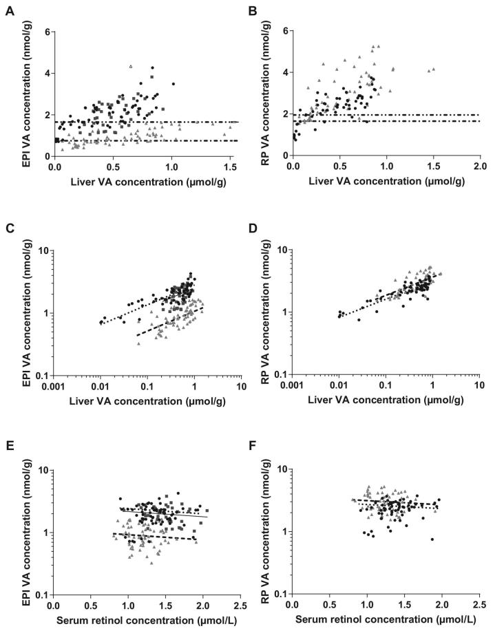
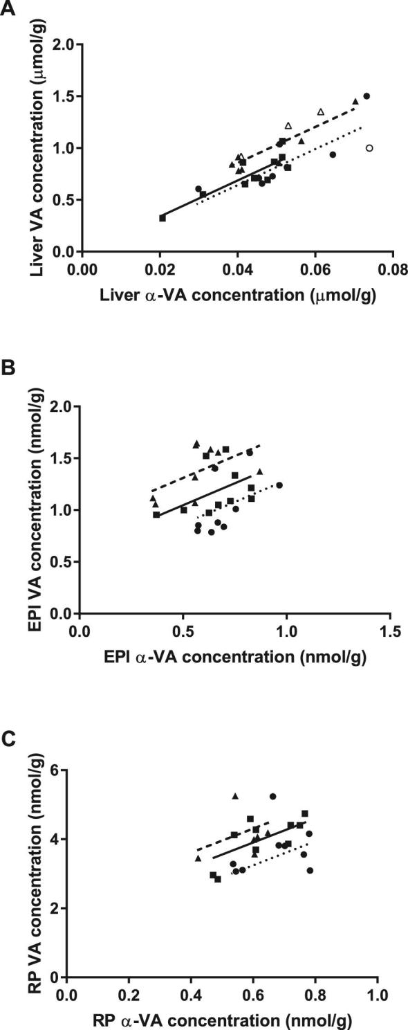
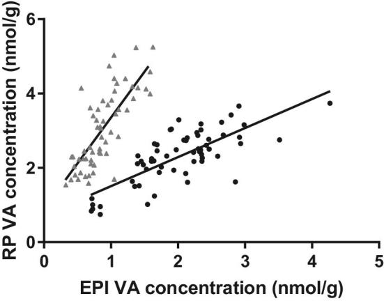
Liver VA did not predict serum retinol concentrations. After logarithmic transformation of adipose and liver values, liver VA positively predicted both adipose depots' VA concentrations ( < 0.001, > 0.7). Adding serum retinol or body fat percentage did not significantly increase the adjusted . In liver, α-VA concentration explained much of the variation of VA ( < 0.001, > 0.7), but far less in epididymal and retroperitoneal adipose ( = 0.004 and 0.012, respectively, < 0.4).
Adipose VA is correlated with liver VA and is a potential biomarker of VA status. It is not fully explained by chylomicron deposition and is negatively affected by serum retinol. Adipose biopsy validation is needed for human applications.
肝脏维生素A(VA)浓度是评估VA状态的金标准,但无法常规获取。脂肪组织中视黄醇和视黄酯形式的VA浓度可能与肝脏VA相关。α-VA(以α-视黄醇形式存在)是α-胡萝卜素的裂解产物,可追踪餐后VA在组织中的分布,但不能从肝脏储存中再循环,有助于了解组织VA的来源。
我们对蒙古沙鼠肝脏和脂肪中的VA和α-VA进行了二次分析,以确定脂肪组织VA作为VA状态生物标志物的适用性。
在3项研究中,186只沙鼠食用含有0-15.9μg(0-55.6nmol)视黄醇活性当量/克的预形成VA和/或维生素A原类胡萝卜素的饲料36-62天。在最后一项研究中通过MRI测定体脂百分比。血清和肝脏中的视黄醇、α-视黄醇和视黄酯经提取后通过HPLC或超高效液相色谱(UPLC)进行分析。附睾和腹膜后脂肪组织中的视黄醇和α-视黄醇在匀浆、皂化和提取后通过UPLC测定。采用适当数据转换的线性回归模型确定脂肪VA浓度的决定因素。
肝脏VA不能预测血清视黄醇浓度。对脂肪和肝脏数据进行对数转换后,肝脏VA可正向预测两个脂肪库的VA浓度(P<0.001,R²>0.7)。加入血清视黄醇或体脂百分比后,调整后的R²没有显著增加。在肝脏中,α-VA浓度解释了VA的大部分变异(P<0.001,R²>0.7),但在附睾和腹膜后脂肪中解释的变异要少得多(分别为P=0.004和0.012,R²<0.4)。
脂肪VA与肝脏VA相关,是VA状态的潜在生物标志物。乳糜微粒沉积不能完全解释其情况,且其受血清视黄醇的负面影响。在人体应用中需要进行脂肪活检验证。