• 文献检索
  • 文档翻译
  • 深度研究
  • 学术资讯
  • Suppr Zotero 插件Zotero 插件
  • 邀请有礼
  • 套餐&价格
  • 历史记录
应用&插件
Suppr Zotero 插件Zotero 插件浏览器插件Mac 客户端Windows 客户端微信小程序
定价
高级版会员购买积分包购买API积分包
服务
文献检索文档翻译深度研究API 文档MCP 服务
关于我们
关于 Suppr公司介绍联系我们用户协议隐私条款
关注我们

Suppr 超能文献

核心技术专利:CN118964589B侵权必究
粤ICP备2023148730 号-1Suppr @ 2026

文献检索

告别复杂PubMed语法,用中文像聊天一样搜索,搜遍4000万医学文献。AI智能推荐,让科研检索更轻松。

立即免费搜索

文件翻译

保留排版,准确专业,支持PDF/Word/PPT等文件格式,支持 12+语言互译。

免费翻译文档

深度研究

AI帮你快速写综述,25分钟生成高质量综述,智能提取关键信息,辅助科研写作。

立即免费体验

自动分割眼科 OCT-A 图像的中心凹无血管区。

Automatic segmentation of the foveal avascular zone in ophthalmological OCT-A images.

机构信息

Department of Computer Science, University of A Coruña, A Coruña, Spain.

CITIC-Research Center of Information and Communication Technologies, University of A Coruña, A Coruña, Spain.

出版信息

PLoS One. 2019 Feb 22;14(2):e0212364. doi: 10.1371/journal.pone.0212364. eCollection 2019.

DOI:10.1371/journal.pone.0212364
PMID:30794594
原文链接:https://pmc.ncbi.nlm.nih.gov/articles/PMC6386246/
Abstract

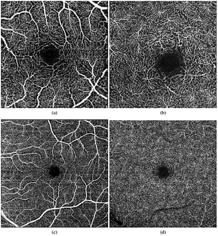
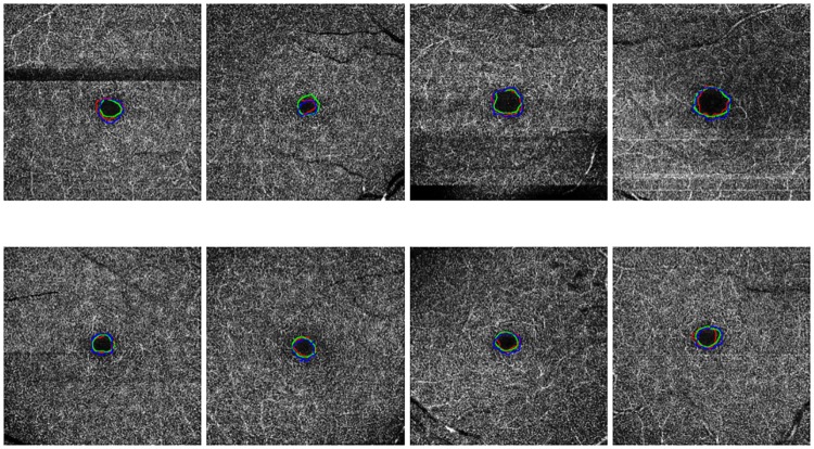
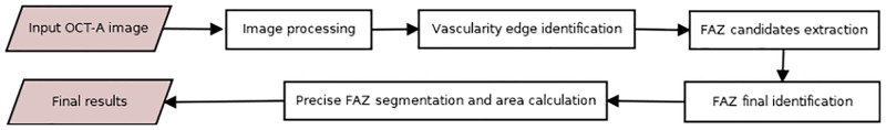
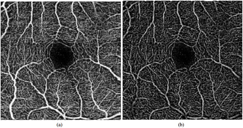
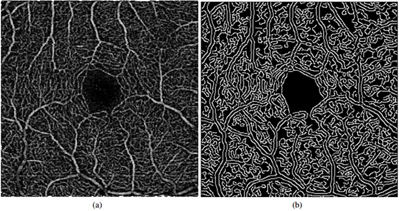
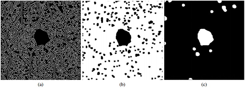
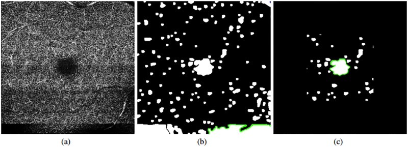
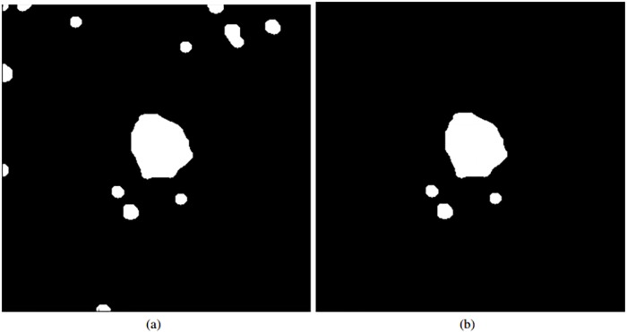
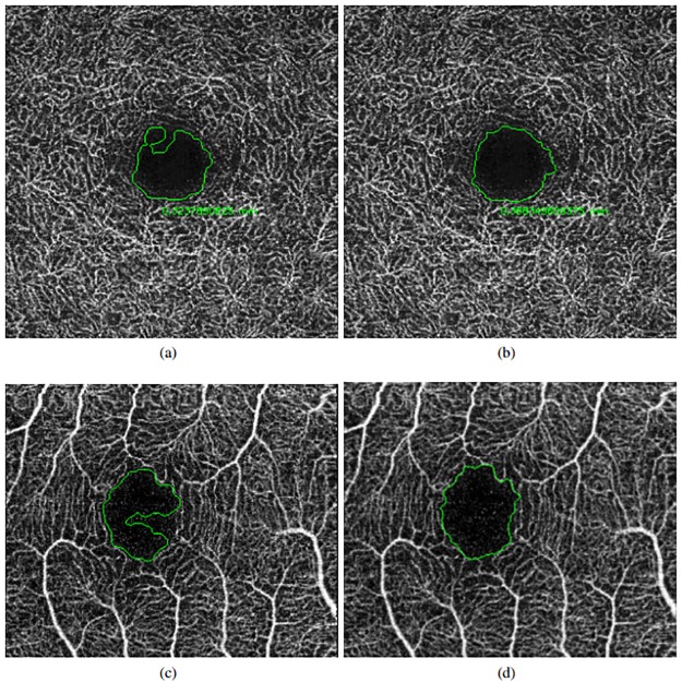
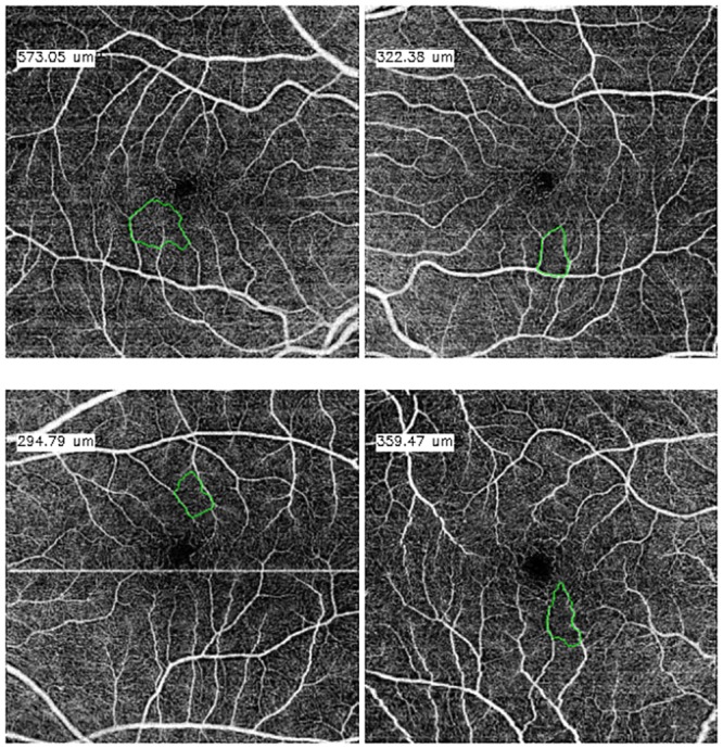
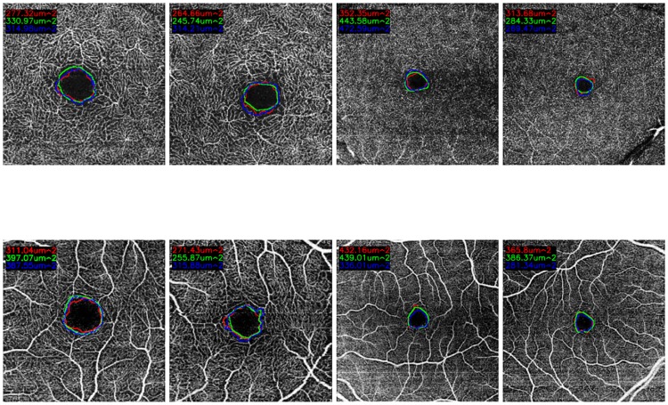
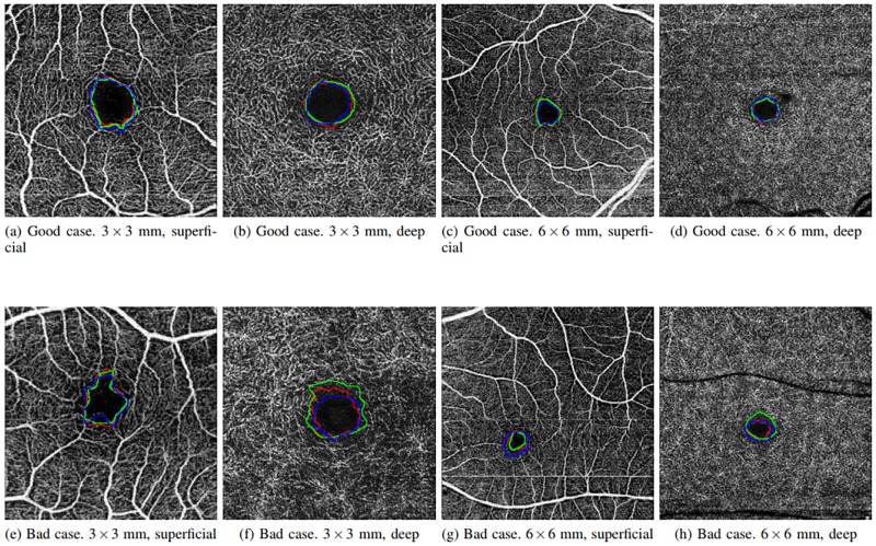
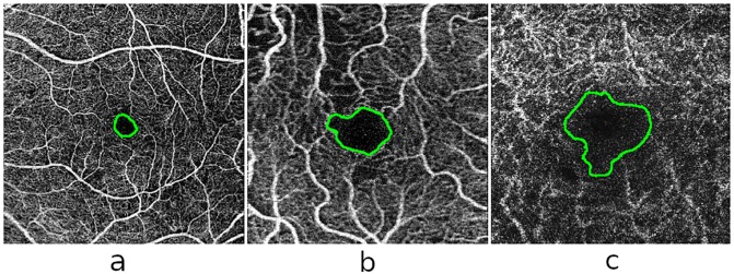
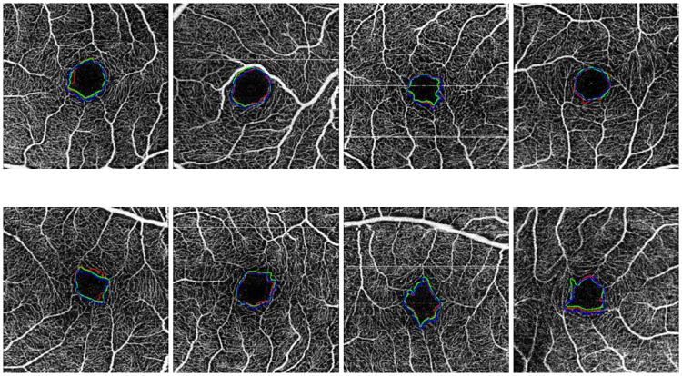
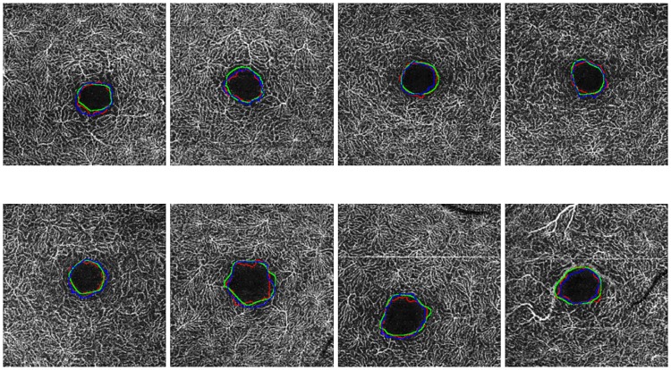
Angiography by Optical Coherence Tomography (OCT-A) is a non-invasive retinal imaging modality of recent appearance that allows the visualization of the vascular structure at predefined depths based on the detection of the blood movement through the retinal vasculature. In this way, OCT-A images constitute a suitable scenario to analyze the retinal vascular properties of regions of interest as is the case of the macular area, measuring the characteristics of the foveal vascular and avascular zones. Extracted parameters of this region can be used as prognostic factors that determine if the patient suffers from certain pathologies (such as diabetic retinopathy or retinal vein occlusion, among others), indicating the associated pathological degree. The manual extraction of these biomedical parameters is a long, tedious and subjective process, introducing a significant intra and inter-expert variability, which penalizes the utility of the measurements. In addition, the absence of tools that automatically facilitate these calculations encourages the creation of computer-aided diagnosis frameworks that ease the doctor's work, increasing their productivity and making viable the use of this type of vascular biomarkers. In this work we propose a fully automatic system that identifies and precisely segments the region of the foveal avascular zone (FAZ) using a novel ophthalmological image modality as is OCT-A. The system combines different image processing techniques to firstly identify the region where the FAZ is contained and, secondly, proceed with the extraction of its precise contour. The system was validated using a representative set of 213 healthy and diabetic OCT-A images, providing accurate results with the best correlation with the manual measurements of two experts clinician of 0.93 as well as a Jaccard's index of 0.82 of the best experimental case in the experiments with healthy OCT-A images. The method also provided satisfactory results in diabetic OCT-A images, with a best correlation coefficient with the manual labeling of an expert clinician of 0.93 and a Jaccard's index of 0.83. This tool provides an accurate FAZ measurement with the desired objectivity and reproducibility, being very useful for the analysis of relevant vascular diseases through the study of the retinal micro-circulation.

摘要

光学相干断层扫描血管造影术(OCT-A)是一种最近出现的非侵入性视网膜成像方式,它允许根据视网膜血管中血液的运动检测,在预设深度可视化血管结构。通过这种方式,OCT-A 图像构成了分析感兴趣区域(如黄斑区域)视网膜血管特性的合适场景,测量黄斑区中心凹血管和无血管区的特征。该区域提取的参数可用作预测因子,以确定患者是否患有某些疾病(如糖尿病性视网膜病变或视网膜静脉阻塞等),并指示相关的病理程度。这些生物医学参数的手动提取是一个漫长、繁琐且主观的过程,引入了显著的专家内和专家间变异性,从而降低了测量的实用性。此外,缺乏自动方便这些计算的工具,鼓励创建计算机辅助诊断框架,减轻医生的工作负担,提高其工作效率,并使这种类型的血管生物标志物的使用成为可能。在这项工作中,我们提出了一种使用新型眼科图像模式(如 OCT-A)自动识别和精确分割中心凹无血管区(FAZ)的全自动系统。该系统结合了不同的图像处理技术,首先识别包含 FAZ 的区域,然后提取其精确轮廓。该系统使用一组有代表性的 213 张健康和糖尿病 OCT-A 图像进行了验证,为最佳实验案例在健康 OCT-A 图像的实验中,与两位临床专家的手动测量结果的相关性最好为 0.93,与手动测量结果的最佳 Jaccard 指数为 0.82,提供了准确的结果。该方法在糖尿病 OCT-A 图像中也取得了令人满意的结果,与一位临床专家的手动标记的最佳相关系数为 0.93,Jaccard 指数为 0.83。该工具提供了准确的 FAZ 测量,具有所需的客观性和可重复性,通过研究视网膜微循环,对相关血管疾病的分析非常有用。

https://cdn.ncbi.nlm.nih.gov/pmc/blobs/da2b/6386246/01be7329e5ac/pone.0212364.g016.jpg
https://cdn.ncbi.nlm.nih.gov/pmc/blobs/da2b/6386246/b30d3ae5b724/pone.0212364.g001.jpg
https://cdn.ncbi.nlm.nih.gov/pmc/blobs/da2b/6386246/49b4e24e1a55/pone.0212364.g002.jpg
https://cdn.ncbi.nlm.nih.gov/pmc/blobs/da2b/6386246/747fb311bfa0/pone.0212364.g003.jpg
https://cdn.ncbi.nlm.nih.gov/pmc/blobs/da2b/6386246/b1619712f2fd/pone.0212364.g004.jpg
https://cdn.ncbi.nlm.nih.gov/pmc/blobs/da2b/6386246/5e765351d852/pone.0212364.g005.jpg
https://cdn.ncbi.nlm.nih.gov/pmc/blobs/da2b/6386246/b8a5216033ec/pone.0212364.g006.jpg
https://cdn.ncbi.nlm.nih.gov/pmc/blobs/da2b/6386246/f732d1cd59cd/pone.0212364.g007.jpg
https://cdn.ncbi.nlm.nih.gov/pmc/blobs/da2b/6386246/5ed3eca1c895/pone.0212364.g008.jpg
https://cdn.ncbi.nlm.nih.gov/pmc/blobs/da2b/6386246/125a581011f5/pone.0212364.g009.jpg
https://cdn.ncbi.nlm.nih.gov/pmc/blobs/da2b/6386246/350a36044e87/pone.0212364.g010.jpg
https://cdn.ncbi.nlm.nih.gov/pmc/blobs/da2b/6386246/3e3b945b06ce/pone.0212364.g011.jpg
https://cdn.ncbi.nlm.nih.gov/pmc/blobs/da2b/6386246/64f85c6a811e/pone.0212364.g012.jpg
https://cdn.ncbi.nlm.nih.gov/pmc/blobs/da2b/6386246/03a29eacd78c/pone.0212364.g013.jpg
https://cdn.ncbi.nlm.nih.gov/pmc/blobs/da2b/6386246/671c98ced278/pone.0212364.g014.jpg
https://cdn.ncbi.nlm.nih.gov/pmc/blobs/da2b/6386246/f695cf2e4fc3/pone.0212364.g015.jpg
https://cdn.ncbi.nlm.nih.gov/pmc/blobs/da2b/6386246/01be7329e5ac/pone.0212364.g016.jpg

相似文献

1
Automatic segmentation of the foveal avascular zone in ophthalmological OCT-A images.自动分割眼科 OCT-A 图像的中心凹无血管区。
PLoS One. 2019 Feb 22;14(2):e0212364. doi: 10.1371/journal.pone.0212364. eCollection 2019.
2
Foveal avascular zone segmentation in optical coherence tomography angiography images using a deep learning approach.利用深度学习方法对光学相干断层扫描血管造影图像中的中心凹无血管区进行分割。
Sci Rep. 2021 Jan 13;11(1):1031. doi: 10.1038/s41598-020-80058-x.
3
Subtle changes in diabetic retinas localised in 3D using OCT.利用光学相干断层扫描(OCT)对糖尿病视网膜的细微变化进行三维定位。
Ophthalmic Physiol Opt. 2018 Sep;38(5):477-491. doi: 10.1111/opo.12578. Epub 2018 Jul 26.
4
Foveal ganglion cell layer damage in ischemic diabetic maculopathy: correlation of optical coherence tomographic and anatomic changes.缺血性糖尿病性黄斑病变中的黄斑神经节细胞层损伤:光学相干断层扫描与解剖学变化的相关性
Ophthalmology. 2009 Oct;116(10):1949-59.e8. doi: 10.1016/j.ophtha.2009.06.066. Epub 2009 Aug 21.
5
Visual Acuity Is Correlated with the Area of the Foveal Avascular Zone in Diabetic Retinopathy and Retinal Vein Occlusion.视力与糖尿病视网膜病变和视网膜静脉阻塞的中心无血管区面积相关。
Ophthalmology. 2016 Nov;123(11):2352-2367. doi: 10.1016/j.ophtha.2016.07.008. Epub 2016 Aug 11.
6
Improved Automated Foveal Avascular Zone Measurement in Cirrus Optical Coherence Tomography Angiography Using the Level Sets Macro.利用水平集宏在 Cirrus 光学相干断层扫描血管造影中进行改进的自动黄斑无血管区测量。
Transl Vis Sci Technol. 2020 Nov 13;9(12):20. doi: 10.1167/tvst.9.12.20. eCollection 2020 Nov.
7
Evaluation of Automatically Quantified Foveal Avascular Zone Metrics for Diagnosis of Diabetic Retinopathy Using Optical Coherence Tomography Angiography.基于光学相干断层扫描血管造影的自动量化中心凹无血管区指标对糖尿病视网膜病变的诊断评估。
Invest Ophthalmol Vis Sci. 2018 May 1;59(6):2212-2221. doi: 10.1167/iovs.17-23498.
8
Noninvasive imaging of the foveal avascular zone with high-speed, phase-variance optical coherence tomography.高速相位变化光学相干断层扫描对黄斑无血管区的无创成像。
Invest Ophthalmol Vis Sci. 2012 Jan 5;53(1):85-92. doi: 10.1167/iovs.11-8249.
9
Optical coherence tomography angiography of the foveal avascular zone in diabetic retinopathy.糖尿病视网膜病变中黄斑无血管区的光学相干断层扫描血管造影
Graefes Arch Clin Exp Ophthalmol. 2016 Jun;254(6):1051-8. doi: 10.1007/s00417-015-3148-2. Epub 2015 Sep 4.
10
Optical Coherence Tomography Angiography Analysis of the Foveal Avascular Zone and Macular Vessel Density After Anti-VEGF Therapy in Eyes With Diabetic Macular Edema and Retinal Vein Occlusion.糖尿病性黄斑水肿和视网膜静脉阻塞患者抗VEGF治疗后黄斑无血管区和黄斑血管密度的光学相干断层扫描血管造影分析
Invest Ophthalmol Vis Sci. 2017 Jan 1;58(1):30-34. doi: 10.1167/iovs.16-20579.

引用本文的文献

1
Real time blood detection in CCTV surveillance using attention enhanced InceptionV3.使用注意力增强型InceptionV3在闭路电视监控中进行实时血液检测。
Sci Rep. 2025 Aug 7;15(1):28977. doi: 10.1038/s41598-025-14941-w.
2
FLA-UNet: feature-location attention U-Net for foveal avascular zone segmentation in OCTA images.FLA-UNet:用于光学相干断层扫描血管造影(OCTA)图像中黄斑无血管区分割的特征位置注意力U-Net
Front Artif Intell. 2025 Jul 17;8:1463233. doi: 10.3389/frai.2025.1463233. eCollection 2025.
3
Low rank based FAZ segmentation in OCTA images.

本文引用的文献

1
Evaluation of Automatically Quantified Foveal Avascular Zone Metrics for Diagnosis of Diabetic Retinopathy Using Optical Coherence Tomography Angiography.基于光学相干断层扫描血管造影的自动量化中心凹无血管区指标对糖尿病视网膜病变的诊断评估。
Invest Ophthalmol Vis Sci. 2018 May 1;59(6):2212-2221. doi: 10.1167/iovs.17-23498.
2
Computer-aided diagnosis of lung nodule using gradient tree boosting and Bayesian optimization.基于梯度提升树和贝叶斯优化的肺结节计算机辅助诊断。
PLoS One. 2018 Apr 19;13(4):e0195875. doi: 10.1371/journal.pone.0195875. eCollection 2018.
3
Enhanced visualization of the retinal vasculature using depth information in OCT.
光学相干断层扫描血管造影(OCTA)图像中基于低秩的黄斑无血管区(FAZ)分割
Sci Rep. 2025 Jul 9;15(1):24757. doi: 10.1038/s41598-025-07448-x.
4
Addressing fairness issues in deep learning-based medical image analysis: a systematic review.解决基于深度学习的医学图像分析中的公平性问题:一项系统综述。
NPJ Digit Med. 2024 Oct 17;7(1):286. doi: 10.1038/s41746-024-01276-5.
5
Foveal avascular zone segmentation using deep learning-driven image-level optimization and fundus photographs.使用深度学习驱动的图像级优化和眼底照片进行黄斑无血管区分割
Proc IEEE Int Symp Biomed Imaging. 2023 Apr;2023. doi: 10.1109/isbi53787.2023.10230410. Epub 2023 Sep 1.
6
Study of Foveal Avascular Zone Growth in Individuals With Mild Diabetic Retinopathy by Optical Coherence Tomography.光学相干断层扫描对轻度糖尿病视网膜病变患者中心凹无血管区生长的研究。
Invest Ophthalmol Vis Sci. 2023 Sep 1;64(12):21. doi: 10.1167/iovs.64.12.21.
7
Enhanced Deep Learning Model for Classification of Retinal Optical Coherence Tomography Images.基于深度学习的视网膜光学相干断层扫描图像分类增强模型。
Sensors (Basel). 2023 Jun 7;23(12):5393. doi: 10.3390/s23125393.
8
The Shape of the Foveal Avascular Zone: When a Circle Isn't Round.黄斑中心凹无血管区的形态:圆非其圆。
Transl Vis Sci Technol. 2023 Jun 1;12(6):26. doi: 10.1167/tvst.12.6.26.
9
A Reliable Criterion for the Correct Delimitation of the Foveal Avascular Zone in Diabetic Patients.糖尿病患者黄斑无血管区正确界定的可靠标准
J Pers Med. 2023 May 12;13(5):822. doi: 10.3390/jpm13050822.
10
The Comparison of Retinal Microvascular Findings in Acute COVID-19 and 1-Year after Hospital Discharge Assessed with Multimodal Imaging-A Prospective Longitudinal Cohort Study.多模态影像评估的急性 COVID-19 与出院后 1 年的视网膜血管变化比较:一项前瞻性纵向队列研究。
Int J Mol Sci. 2023 Feb 17;24(4):4032. doi: 10.3390/ijms24044032.
利用 OCT 中的深度信息增强视网膜血管可视化。
Med Biol Eng Comput. 2017 Dec;55(12):2209-2225. doi: 10.1007/s11517-017-1660-8. Epub 2017 Jun 17.
4
Hydra: A web-based system for cardiovascular analysis, diagnosis and treatment.九头蛇:一个基于网络的心血管分析、诊断和治疗系统。
Comput Methods Programs Biomed. 2017 Feb;139:61-81. doi: 10.1016/j.cmpb.2016.10.019. Epub 2016 Oct 29.
5
Optical Coherence Tomography Angiography Findings in Stargardt Disease.斯塔加特病的光学相干断层扫描血管造影结果
PLoS One. 2017 Feb 2;12(2):e0170343. doi: 10.1371/journal.pone.0170343. eCollection 2017.
6
A review of optical coherence tomography angiography (OCTA).光学相干断层扫描血管造影术(OCTA)综述。
Int J Retina Vitreous. 2015 Apr 15;1:5. doi: 10.1186/s40942-015-0005-8. eCollection 2015.
7
Reproducibility and repeatability of foveal avascular zone area measurements using swept-source optical coherence tomography angiography in healthy subjects.在健康受试者中使用扫频源光学相干断层扫描血管造影术测量黄斑无血管区面积的可重复性和重复性
Eur J Ophthalmol. 2017 May 11;27(3):336-341. doi: 10.5301/ejo.5000858. Epub 2016 Sep 9.
8
Visual Acuity Is Correlated with the Area of the Foveal Avascular Zone in Diabetic Retinopathy and Retinal Vein Occlusion.视力与糖尿病视网膜病变和视网膜静脉阻塞的中心无血管区面积相关。
Ophthalmology. 2016 Nov;123(11):2352-2367. doi: 10.1016/j.ophtha.2016.07.008. Epub 2016 Aug 11.
9
Automated Quantification of Capillary Nonperfusion Using Optical Coherence Tomography Angiography in Diabetic Retinopathy.利用光学相干断层扫描血管造影术自动定量分析糖尿病视网膜病变中的毛细血管无灌注情况。
JAMA Ophthalmol. 2016 Apr;134(4):367-73. doi: 10.1001/jamaophthalmol.2015.5658.
10
Reproducibility and repeatability of foveal avascular zone measurements in healthy subjects by optical coherence tomography angiography.光学相干断层扫描血管造影术测量健康受试者黄斑无血管区的可重复性和重复性
Br J Ophthalmol. 2016 May;100(5):671-6. doi: 10.1136/bjophthalmol-2015-307330. Epub 2015 Sep 16.