Proteomics program, Novo Nordisk Foundation Center for Protein Research, Faculty of Health and Medical Sciences, University of Copenhagen, Blegdamsvej 3B, 2200 Copenhagen, Denmark.
Proteomics program, Novo Nordisk Foundation Center for Protein Research, Faculty of Health and Medical Sciences, University of Copenhagen, Blegdamsvej 3B, 2200 Copenhagen, Denmark..
Mol Cell Proteomics. 2019 May;18(5):1010-1026. doi: 10.1074/mcp.TIR119.001315. Epub 2019 Feb 23.
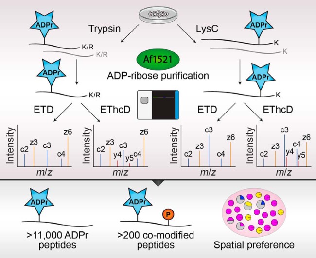
ADP-ribosylation is a widespread post-translational modification (PTM) with crucial functions in many cellular processes. Here, we describe an in-depth ADP-ribosylome using our Af1521-based proteomics methodology for comprehensive profiling of ADP-ribosylation sites, by systematically assessing complementary proteolytic digestions and precursor fragmentation through application of electron-transfer higher-energy collisional dissociation (EThcD) and electron transfer dissociation (ETD), respectively. Although ETD spectra yielded higher identification scores, EThcD generally proved superior to ETD in identification and localization of ADP-ribosylation sites regardless of protease employed. Notwithstanding, the propensities of complementary proteases and fragmentation methods expanded the detectable repertoire of ADP-ribosylation to an unprecedented depth. This system-wide profiling of the ADP-ribosylome in HeLa cells subjected to DNA damage uncovered >11,000 unique ADP-ribosylated peptides mapping to >7,000 ADP-ribosylation sites, in total modifying over one-third of the human nuclear proteome and highlighting the vast scope of this PTM. High-resolution MS/MS spectra enabled identification of dozens of proteins concomitantly modified by ADP-ribosylation and phosphorylation, revealing a considerable degree of crosstalk on histones. ADP-ribosylation was confidently localized to various amino acid residue types, including less abundantly modified residues, with hundreds of ADP-ribosylation sites pinpointed on histidine, arginine, and tyrosine residues. Functional enrichment analysis suggested modification of these specific residue types is directed in a spatial manner, with tyrosine ADP-ribosylation linked to the ribosome, arginine ADP-ribosylation linked to the endoplasmic reticulum, and histidine ADP-ribosylation linked to the mitochondrion.
ADP-核糖基化是一种广泛存在的翻译后修饰(PTM),在许多细胞过程中具有关键功能。在这里,我们使用基于 Af1521 的蛋白质组学方法描述了一个深入的 ADP-核糖基组,该方法通过系统评估互补的蛋白水解消化和通过应用电子转移更高能量碰撞解离(EThcD)和电子转移解离(ETD)分别对前体片段化进行全面的 ADP-核糖基化位点分析。尽管 ETD 光谱产生了更高的鉴定分数,但无论使用哪种蛋白酶,EThcD 通常在鉴定和定位 ADP-核糖基化位点方面都优于 ETD。尽管如此,互补蛋白酶和片段化方法的倾向扩展了 ADP-核糖基化的可检测范围,达到了前所未有的深度。在 DNA 损伤下用 HeLa 细胞进行的 ADP-核糖基组的系统分析揭示了 >11000 个独特的 ADP-核糖基化肽,映射到 >7000 个 ADP-核糖基化位点,总共修饰了超过三分之一的人类核蛋白质组,并突出了这种 PTM 的巨大范围。高分辨率 MS/MS 光谱能够鉴定出数十种同时被 ADP-核糖基化和磷酸化修饰的蛋白质,揭示了组蛋白上相当程度的串扰。ADP-核糖基化被自信地定位到各种氨基酸残基类型,包括修饰程度较低的残基,在组氨酸、精氨酸和酪氨酸残基上有数百个 ADP-核糖基化位点。功能富集分析表明,这些特定残基类型的修饰以空间方式进行,酪氨酸 ADP-核糖基化与核糖体相关,精氨酸 ADP-核糖基化与内质网相关,组氨酸 ADP-核糖基化与线粒体相关。