1 Department of Physiology, Saga Medical School, Saga, Japan.
2 Department of Anesthesiology & Critical Care Medicine, Saga Medical School, Saga, Japan.
Mol Pain. 2019 Jan-Dec;15:1744806918824243. doi: 10.1177/1744806918824243.
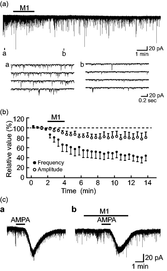
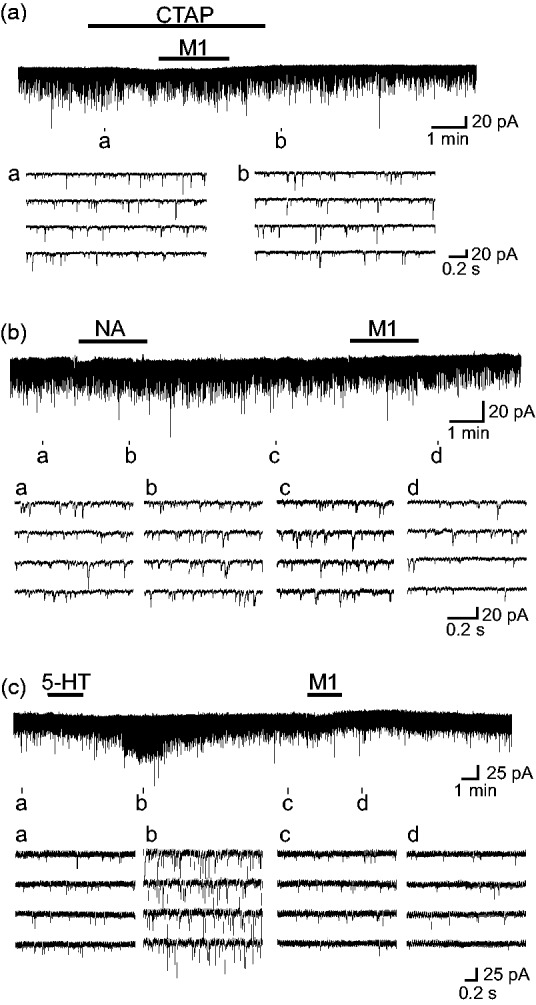
To reveal cellular mechanisms for antinociception produced by clinically used tramadol, we investigated the effect of its metabolite O-desmethyltramadol (M1) on glutamatergic excitatory transmission in spinal dorsal horn lamina II (substantia gelatinosa; SG) neurons. The whole-cell patch-clamp technique was applied at a holding potential of -70 mV to SG neurons of an adult rat spinal cord slice with an attached dorsal root. Under the condition where a postsynaptic action of M1 was inhibited, M1 superfused for 2 min reduced the frequency of spontaneous excitatory postsynaptic current in a manner sensitive to a μ-opioid receptor antagonist CTAP; its amplitude and also a response of SG neurons to bath-applied AMPA were hardly affected. The presynaptic effect of M1 was different from that of noradrenaline or serotonin which was examined in the same neuron. M1 also reduced by almost the same extent the peak amplitudes of monosynaptic primary-afferent Aδ-fiber and C-fiber excitatory postsynaptic currents evoked by stimulating the dorsal root. These actions of M1 persisted for >10 min after its washout. These results indicate that M1 inhibits the quantal release of L-glutamate from nerve terminals by activating μ-opioid but not noradrenaline and serotonin receptors; this inhibition is comparable in extent between monosynaptic primary-afferent Aδ-fiber and C-fiber transmissions. Considering that the SG plays a pivotal role in regulating nociceptive transmission, the present findings could contribute to at least a part of the inhibitory action of tramadol on nociceptive transmission together with its hyperpolarizing effect as reported previously.
为了揭示临床应用的曲马多产生的抗伤害作用的细胞机制,我们研究了其代谢产物 O-去甲曲马多(M1)对脊髓背角 II 层(胶状质;SG)神经元谷氨酸能兴奋性传递的影响。在成年大鼠脊髓切片上附有背根的情况下,应用全细胞膜片钳技术,在-70 mV 的保持电位下,对 SG 神经元进行检测。在抑制 M1 的突触后作用的条件下,2 分钟内持续给予 M1 可降低自发性兴奋性突触后电流的频率,其方式对μ-阿片受体拮抗剂 CTAP 敏感;其幅度以及 SG 神经元对浴中应用 AMPA 的反应几乎不受影响。M1 的突触前作用不同于在同一神经元中检测到的去甲肾上腺素或 5-羟色胺。M1 还几乎同等程度地减少了刺激背根时诱发的单突触初级传入 Aδ 纤维和 C 纤维兴奋性突触后电流的峰值幅度。M1 的这些作用在其洗脱后持续>10 分钟。这些结果表明,M1 通过激活μ-阿片受体而不是去甲肾上腺素和 5-羟色胺受体,抑制来自神经末梢的 L-谷氨酸的量子释放;这种抑制在单突触初级传入 Aδ 纤维和 C 纤维传递之间的程度相当。考虑到 SG 在调节伤害性传递中起着关键作用,本研究结果可能有助于至少部分解释曲马多对伤害性传递的抑制作用,以及其先前报道的超极化作用。