Department of Diagnostic and Interventional Radiology, Technical University of Munich, Munich, Germany.
Department of Diagnostic and Interventional Neuroradiology, Technical University of Munich, Munich, Germany.
PLoS One. 2019 Feb 25;14(2):e0212679. doi: 10.1371/journal.pone.0212679. eCollection 2019.
To evaluate the accuracy of Spectral Photon-Counting Computed Tomography (SPCCT) in the quantification of iodine concentrations and its potential for the differentiation between blood and iodine.
Tubes with blood and a concentration series of iodine were scanned with a preclinical SPCCT system (both in vitro and in an ex vivo bovine brain tissue sample). Iodine density maps (IDM) and virtual non-contrast (VNC) images were generated using the multi-bin spectral information to perform material decomposition. Region-of-interest (ROI) analysis was performed within the tubes to quantitatively determine the absolute content of iodine (mg/ml).
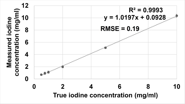
In conventional CT images, ROI analysis showed similar Hounsfield Unit (HU) values for the tubes with blood and iodine (59.9 ± 1.8 versus 59.2 ± 1.5). Iodine density maps enabled clear differentiation between blood and iodine in vitro, as well as in the bovine brain model. Quantitative measurements of the different iodine concentrations matched well with those of actual known concentrations even for very small iodine concentrations with values below 1mg/ml (RMSE = 0.19).
SPCCT providing iodine maps and virtual non-contrast images allows material decomposition, differentiation between blood and iodine in vitro and ex vivo in a bovine brain model and reliably quantifies the iodine concentration.
评估光谱光子计数 CT(SPCCT)在碘浓度定量中的准确性及其用于区分血液和碘的潜力。
使用临床前 SPCCT 系统对具有血液和碘浓度系列的管进行扫描(均进行体外和牛脑组织样本的离体扫描)。使用多-bin 光谱信息生成碘密度图(IDM)和虚拟非对比(VNC)图像,以进行物质分解。在管内进行感兴趣区域(ROI)分析,以定量确定碘的绝对含量(mg/ml)。
在常规 CT 图像中,ROI 分析显示血液和碘管的 Hounsfield 单位(HU)值相似(59.9 ± 1.8 与 59.2 ± 1.5)。碘密度图能够在体外以及牛脑模型中清楚地区分血液和碘。即使对于非常小的碘浓度(低于 1mg/ml),不同碘浓度的定量测量也与实际已知浓度非常吻合(RMSE = 0.19)。
SPCCT 提供碘图和虚拟非对比图像,允许在体外和牛脑模型中进行物质分解、血液和碘的区分,并可靠地定量碘浓度。