Rukmini A V, Milea Dan, Gooley Joshua J
Programme in Neuroscience and Behavioural Disorders, Centre for Cognitive Neuroscience, Duke-NUS Medical School, Singapore, Singapore.
Singapore National Eye Centre, Singapore Eye Research Institute, Singapore, Singapore.
Front Neurol. 2019 Feb 12;10:76. doi: 10.3389/fneur.2019.00076. eCollection 2019.
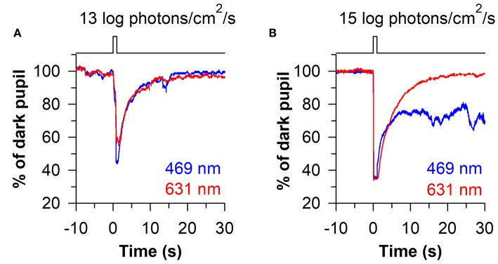
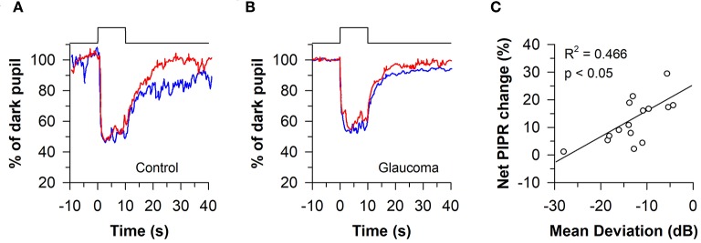
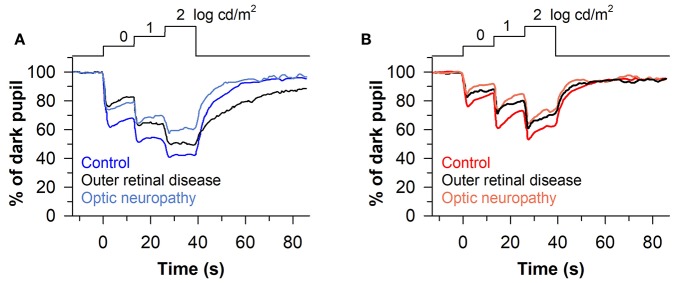
The pupillary light reflex is mediated by melanopsin-containing intrinsically-photosensitive retinal ganglion cells (ipRGCs), which also receive input from rods and cones. Melanopsin-dependent pupillary light responses are short-wavelength sensitive, have a higher threshold of activation, and are much slower to activate and de-activate compared with rod/cone-mediated responses. Given that rod/cone photoreceptors and melanopsin differ in their response properties, light stimuli can be designed to stimulate preferentially each of the different photoreceptor types, providing a read-out of their function. This has given rise to chromatic pupillometry methods that aim to assess the health of outer retinal photoreceptors and ipRGCs by measuring pupillary responses to blue or red light stimuli. Here, we review different types of chromatic pupillometry protocols that have been tested in patients with retinal or optic nerve disease, including approaches that use short-duration light exposures or continuous exposure to light. Across different protocols, patients with outer retinal disease (e.g., retinitis pigmentosa or Leber congenital amaurosis) show reduced or absent pupillary responses to dim blue-light stimuli used to assess rod function, and reduced responses to moderately-bright red-light stimuli used to assess cone function. By comparison, patients with optic nerve disease (e.g., glaucoma or ischemic optic neuropathy, but not mitochondrial disease) show impaired pupillary responses during continuous exposure to bright blue-light stimuli, and a reduced post-illumination pupillary response after light offset, used to assess melanopsin function. These proof-of-concept studies demonstrate that chromatic pupillometry methods can be used to assess damage to rod/cone photoreceptors and ipRGCs. In future studies, it will be important to determine whether chromatic pupillometry methods can be used for screening and early detection of retinal and optic nerve diseases. Such methods may also prove useful for objectively evaluating the degree of recovery to ipRGC function in blind patients who undergo gene therapy or other treatments to restore vision.
瞳孔光反射由含黑视蛋白的内在光敏视网膜神经节细胞(ipRGCs)介导,这些细胞也接收来自视杆细胞和视锥细胞的输入。依赖黑视蛋白的瞳孔光反应对短波长敏感,激活阈值较高,与视杆/视锥介导的反应相比,激活和失活速度要慢得多。鉴于视杆/视锥光感受器和黑视蛋白的反应特性不同,可以设计光刺激来优先刺激每种不同的光感受器类型,从而读出它们的功能。这催生了色觉瞳孔测量法,旨在通过测量瞳孔对蓝光或红光刺激的反应来评估视网膜外层光感受器和ipRGCs的健康状况。在此,我们回顾了已在视网膜或视神经疾病患者中测试的不同类型的色觉瞳孔测量方案,包括使用短时间光暴露或持续光暴露的方法。在不同的方案中,患有视网膜外层疾病(如色素性视网膜炎或莱伯先天性黑蒙)的患者对用于评估视杆功能的暗光蓝光刺激的瞳孔反应减弱或消失,对用于评估视锥功能的中度明亮红光刺激的反应也减弱。相比之下,患有视神经疾病(如青光眼或缺血性视神经病变,但不包括线粒体疾病)的患者在持续暴露于明亮蓝光刺激期间瞳孔反应受损,并且在光熄灭后用于评估黑视蛋白功能的光照后瞳孔反应减弱。这些概念验证研究表明,色觉瞳孔测量法可用于评估视杆/视锥光感受器和ipRGCs的损伤。在未来的研究中,确定色觉瞳孔测量法是否可用于视网膜和视神经疾病的筛查和早期检测将很重要。这些方法也可能被证明有助于客观评估接受基因治疗或其他恢复视力治疗的盲人患者ipRGC功能的恢复程度。